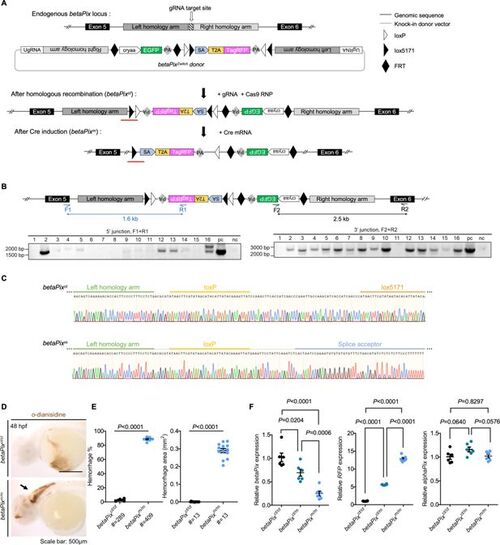
Generation of betaPix conditional trap (betaPixct) allele by a homologous recombination (HDR)-mediated knock-in method. (A) Schematic diagram illustrates the HDR-mediated Zwitch strategy for generating zebrafish knock-in allele at the betaPix locus. (B) Genomic PCR analysis of the F1 embryos confirming the right Zwitch insertions. (C) Sanger sequencing confirming the junction of betaPixct (after HDR-mediated insertion) or betaPixm (after Cre-mediated inversion) that are highlighted by red lines in (A). (D) Representative stereomicroscopy images of erythrocytes stained with o-dianisidine in betaPixct/ct and betaPixm/m embryos at 48 hpf. Brain hemorrhages, indicated with an arrow, in Cre mRNA-injected embryos (betaPixm/m). Lateral views, anterior to the left. (E) Quantification of hemorrhagic parameters in (D). Left panel showing hemorrhage percentages, with independent experiments as dots. Right panel showing hemorrhage areas, with each dot representing one embryo, # represents the numbers of embryos scored for each analysis, and three or more individual experiments conducted. Data are presented in mean ± SEM; unpaired Student’s t-test with individual p-values mentioned in the figure. (F) qRT-PCR analysis showing the expression of betaPix, RFP, and alphaPix in betaPixct/ct, betaPixct/m, and betaPixm/m embryos at 48 hpf. Each dot represents one embryo. Data are presented in mean ± SEM; one-way ANOVA with Dunnett’s test, individual p-values mentioned in the figure. cryaa, αA-crystallin; PA, polyadenylation signal; SA, splice acceptor; T2A, T2A self-cleaving peptide. Individual scale bars are indicated in the figure.
|
