|
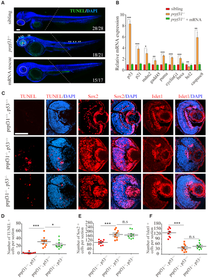
Increased apoptosis and activated p53 pathway in RPCs of prpf31−/− zebrafish. (A) TUNEL staining showed numerous apoptotic cells in the retina, brain and posterior segment of spinal cord in prpf31 mutants at 48 hpf. Injection of wild-type prpf31 mRNAs reduced the apoptotic cells to a normal level. Dotted lines indicate the boundary of two images from the same embryo. White arrows, apoptotic signals. Scale bar, 100 μm. (B) The up-regulation of p53 pathway genes in prpf31 mutants at 36 hpf as detected by qPCR. (C) Deletion of p53 in prpf31 mutants significantly reduced cell apoptosis, but could not rescue the differentiation defects of RPCs. Scale bar, 100 μm. (D−F) The quantitative analysis of TUNEL positive cells, Sox2 positive cells and Islet1 positive cells shown in (C). n = 9 for each panel. Scale bar, 100 μm.
|