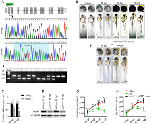
CRISPR/Cas9-mediated knockout of prpf31 led to retinal morphological defects. (A) The gene structure of prpf31 and CRISPR/Cas9 target site were shown. DNA sequencing of the corresponding genomic region revealed a 134 bp deletion/18 bp insertion mutation. (B) Genotype validation by DNA electrophoresis. +/+, wild-type sibling; +/−, heterozygotes; −/−, homozygotes. (C) Relative expression of prpf31 mRNA was detected by qPCR at 36 and 48 hpf. (D) The protein level of Prpf31 was decreased in prpf31−/− embryos at 36 and 48 hpf as detected by western blot. GAPDH was used as an internal control. (E) The morphology of bodies and eyes in prpf31−/− embryos. No obvious defects were observed until 48 hpf. At 60 hpf, the prpf31−/− embryos showed microphthalmia, smaller head and curved body axis. At 72 hpf, these phenotypes were further aggravated. (F) Injection of zebrafish prpf31 mRNA could rescue the developmental defects of mutant zebrafish. (G, H) Quantification of the embryonic length and eye size in the wild-type, prpf31−/− and prpf31−/− + mRNA rescued embryos at 36, 48, 60 and 72 hpf. n = 20 for each panel.
|
