
Figure 3
- ID
- ZDB-IMAGE-210307-99
- Genes
- Antibodies
- Publication
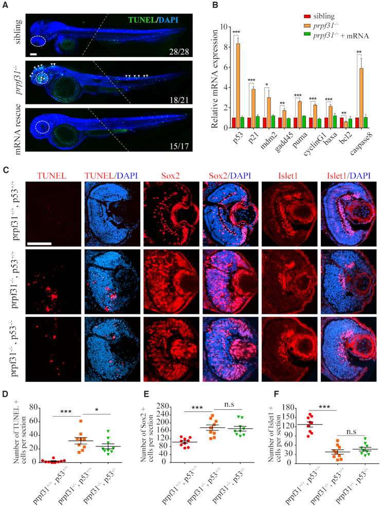
- Li et al., 2021 - Prpf31 is essential for the survival and differentiation of retinal progenitor cells by modulating alternative splicing
- All Figures
- Figures for Li et al., 2021
|
Figure 3
Increased apoptosis and activated