Fig. 1
|
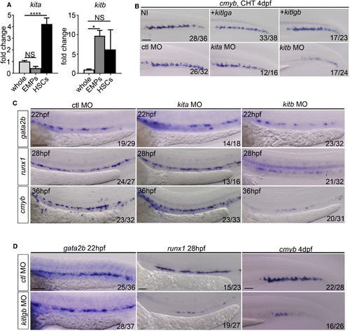
kitb, but Not kita, Signaling Is Necessary for Correct HSC Specification (A) qPCR expression of kita and kitb in FACS-sorted hematopoietic progenitors. Kita whole and EMPS, p = 0.14. kita whole and HSCs, p < 0.0001. kitb whole and EMPs, p = 0.023. kitb whole and HSCs, p = 0.14. EMPs: double-positive lmo2:eGFP, gata1:DsRed cells (28 hpf) (Bertrand et al., 2007). HSCs: double-positive flk1:mCherry, cmyb:eGFP cells (36 hpf) (Bertrand et al., 2010). Data are from biological triplicates. Data are means ± SD. (B) cmyb ISH at 4dpf in non-injected control (NI), kitlga (injected at 500 pg throughout, unless stated) or kitlgb (injected at 200 pg throughout, unless stated) mRNA injected embryos. cmyb was socred at 4dpf following ctrl-MO, kita-MO (injectedat 3 ng throughout, unless stated) or kitb-MO (injected at 8 ng throughout, unless stated) injection. NI, non-injected control. +kitlga/kitlgb, kitlga/kitlgb full length mRNA injected embryos. (C) Control-MO, kita-MO and kitb-MO injected embryos were scored for the expression of gata2b, runx1 and cmyb at 22 hpf, 28 hpf and 36 hpf, respectively. (D) kitlgb morphants were scored for gata2b, runx1 and cmyb at 22 hpf, 28 hpf and 4 dpf, respectively. Analysis was completed using ordinary one way ANOVA with multiple comparisons. **** p < 0.0001; *p < 0.05; NS, p > 0.05. All scale bars, 100 µm. |
| Genes: | |
|---|---|
| Fish: | |
| Knockdown Reagents: | |
| Anatomical Term: | |
| Stage Range: | 26+ somites to Day 4 |
| Fish: | |
|---|---|
| Knockdown Reagents: | |
| Observed In: | |
| Stage Range: | Prim-5 to Day 4 |

