|
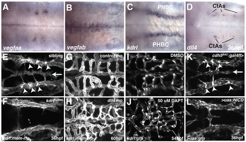
VegfA and Notch signaling are required for angiogenic sprouting in the zebrafish hindbrain. (A-D) Whole-mount in situ hybridization showing the expression of vegfaa (A), vegfab (B), kdrl (C) and dll4 (D) at 36 hpf. (E-L) Maximal intensity projections of confocal z-stacks at 36 (E,F,K,L), 54 (I,J) or 60 (G,H) hpf. Arrowheads indicate CtA sprouts emerging from the PHBC. Arrow indicates the position of the BA. (E,F) Sibling (E) or kdrl mutant (F) embryos, with endothelial cells labeled by kdrl:mem-rfp expression. (G,H) kdrl:mem-rfp-expressing embryos injected with control MO (G) or dll4 MO (H). (I,J) kdrl:gfp-expressing embryos treated from 30 to 54 hpf with DMSO (I) or DAPT (J). (K,L) Embryos from a cross between cdh5:gal4ff, uas:gfp and uas:NICD. The embryo in K is negative for uas:NICD, whereas that in L is heterozygous for uas:NICD. Endothelial cells are labeled by cdh5:gal4ff, uas:gfp expression. Dorsal views, anterior to the left.
|