
Fig. S3
- ID
- ZDB-FIG-110428-18
- Publication
- Bussmann et al., 2011 - Arterial-venous network formation during brain vascularization involves hemodynamic regulation of chemokine signaling
- Other Figures
- All Figure Page
- Back to All Figure Page
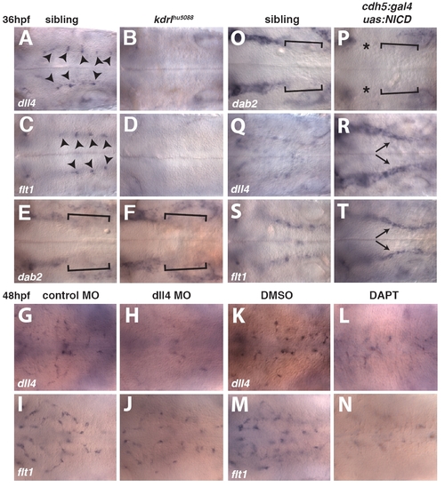
Gene expression changes in kdrlhθ5088 mutant embryos and embryos with increased or decreased Notch signaling pathway activation. (A-F) Whole-mount in situ hybridization showing the expression of dll4 (A,B), flt1 (C,D) and dab2 (E,F) at 36 hpf in sibling control (A,C,E) and kdrlhu5088 mutant (B,D,F) embryos. Optical section dorsal to the PHBC (A-D) or at the level of the PHBC (E,F). Arrowheads indicate dll4 (A) and flt1 (C) expression in CtA sprouts. Brackets indicate reduced expression of dab2 in the posterior PHBC (E,F). (G-J) Whole-mount in situ hybridization showing the expression of dll4 (G,H) or flt1 (I,J) in control MO-injected (G,I) or dll4 MO-injected (H,J) embryos. (K-N) Whole-mount in situ hybridization showing the expression of dll4 (K,L) or flt1 (M,N) in embryos treated with 0.2% DMSO between 30 hpf and 48 hpf (K,M) and embryos treated with 50 μM DAPT between 30 hpf and 48 hpf (L,N). (O-T) Whole-mount in situ hybridization showing the distribution of dab2 (O,P), dll4 (Q,R) and flt1 (S,T) in sibling (O,Q,S) or cdh5:gal4ff; uas:NICD double-transgenic (P,R,T) embryos at 36 hpf, optical sections at the level of the PHBC. Brackets indicate reduced expression of dab2 in the posterior PHBC (O,P). Asterisks mark reduced dab2 expression in the anterior PHBCs in cdh5:gal4ff; uas:NICD double-transgenic embryos (P). Arrows indicate ectopic expression of dll4 (R) and flt1 (T) throughout the PHBC in cdh5:gal4ff; uas:NICD double-transgenic embryos. |