
Fig. 5
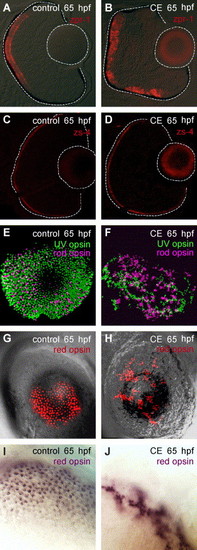
Photoreceptor differentiation and planar organization in control and γ-secretase-inhibited (CE) embryos. (A, B) Double-cones labeled by zpr-1 at 65 hpf (N = 15 embryos). (C, D) The outer limiting membrane (apical surface) was labeled by zs-4 (N = 15 embryos). (E–J) Whole-mount preparations of control and CE-treated eyes processed for in situ hybridization using opsin-specific riboprobes. Rod (Fast Red-pseudo-colored magenta) and UV (FITC-green) opsin in control (E) and CE-treated (F) retinas at 65 hpf. Red opsin (Fast Red-red) in control (G) and CE-treated (H) retinas at 65 hpf. Higher magnification of control (I) and CE-treated (J) retinas stained for red opsin expression (NBT/BCIP-purple). Scale bar = 50 μm (A-H); 20 μm (I, J). |
| Genes: | |
|---|---|
| Fish: | |
| Condition: | |
| Anatomical Term: | |
| Stage: | Pec-fin |
Reprinted from Developmental Biology, 278(2), Bernardos, R.L., Lentz, S.I., Wolfe, M.S., and Raymond, P.A., Notch-Delta signaling is required for spatial patterning and Muller glia differentiation in the zebrafish retina, 381-395, Copyright (2005) with permission from Elsevier. Full text @ Dev. Biol.