
FIGURE
Fig. 8
Fig. 8
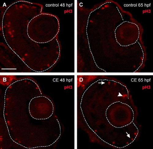
Mitotic activity in the retinas of control and γ-secretase-inhibited (CE) embryos. (A–D) Retinal cryosections labeled with phosphorylated histone H3 (pH3) antibody (Cy3—red). (A) Control and (B) CE-treated retinas labeled with anti-pH3 at 48 hpf. Mitotic cells were present in the outer retina in control and drug-treated retinas. By 65 hpf, mitotic activity was restricted to the germinal zone at the ciliary margin (arrow) and outer nuclear layer (arrowheads) in control (C) and CE-treated (D). Scale bar = 50 μm (A–D). |
Expression Data
Expression Detail
Antibody Labeling
Phenotype Data
Phenotype Detail
Acknowledgments
This image is the copyrighted work of the attributed author or publisher, and
ZFIN has permission only to display this image to its users.
Additional permissions should be obtained from the applicable author or publisher of the image.
Reprinted from Developmental Biology, 278(2), Bernardos, R.L., Lentz, S.I., Wolfe, M.S., and Raymond, P.A., Notch-Delta signaling is required for spatial patterning and Muller glia differentiation in the zebrafish retina, 381-395, Copyright (2005) with permission from Elsevier. Full text @ Dev. Biol.