
Fig. S1
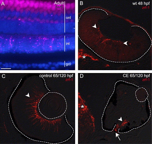
(A) Expression of her6/hes1 (pseudo-colored magenta) in an adult wild-type zebrafish retina co-labeled with DAPI (blue). her6/hes1 expression is present in the inner nuclear layer (inl), and a few cells in the outer nuclear layer (onl), but is absent from the ganglion cell layer (gcl). The apparent labeling in the onl is due to photoreceptor autofluorescence. (B) Expression of zrf-1/GFAP in the retina (arrowheads) and brain (*) of a wild-type embryo at 48 hpf. The earliest expression of GFAP in zebrafish Müller glia reported previously was 5 dpf (Scheer et al., 2001). (C, D) Retinas from embryos treated in control and CE solutions up to 65 hpf and then transferred to drug-free embryo media to continue development up to 120 hpf. At 120 hpf in a control retina (C) zrf-1/GFAP-labeled profiles (arrowhead) span the retina, whereas in a γ-secretase-inhibited (CE) embryo (D), there is no staining in the inner retina, but staining is present (arrowhead) at the optic disc (arrow) and in the brain (*) (N = 15 embryos). Scale bar = 15 μm (A), 50 μm (C, D) or 30 μm (B). |
Reprinted from Developmental Biology, 278(2), Bernardos, R.L., Lentz, S.I., Wolfe, M.S., and Raymond, P.A., Notch-Delta signaling is required for spatial patterning and Muller glia differentiation in the zebrafish retina, 381-395, Copyright (2005) with permission from Elsevier. Full text @ Dev. Biol.