Fig. 6
|
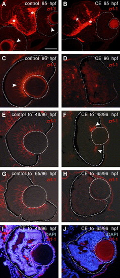
Müller glia fail to differentiate in the absence of Notch–Delta signaling. Immunostaining with zrf-1/GFAP (Cy3—red). In a control embryo at 65 hpf (A), radial glia in the brain are marked with an asterisk (*) and in the retina, endfeet of Müller glia at the vitreal surface are indicated by arrowheads. The apparent immunoreactivity in the lens is non-specific background. In an embryo treated in γ-secretase inhibitor (CE) up to 65 hpf (B), there is no staining with zrf-1/GFAP in the retina (outlined), but zrf-1/GFAP is present in the brain (*) (N = 20 embryos). At 96 hpf in a control retina (C) zrf-1/GFAP-labeled profiles (arrowhead) span the retina, whereas in a CE-treated embryo (D), there is no staining in the retina (outlined) (N = 20 embryos). (E, F) Retinas from embryos treated in control or drug solutions up to 48 hpf and then transferred to drug-free embryo media to continue development up to 96 hpf. In a CE-treated embryo (F) (N = 20 embryos examined), a few Müller glia (arrowheads) are indicated at the periphery of the retina (outlined) in contrast to the numerous Müller glia present in the control retina (E). (G, H) Retinas from embryos treated in control or drug solutions up to 65 hpf and then transferred to drug-free embryo media to continue development up to 96 hpf. In a drug-treated embryo (H), no zrf-1/GFAP staining is present in the retina (outlined) (N = 20 embryos). (I, J) Overlays of DAPI-stained and zrf-1/GFAP images show the relationship of Müller glia to the laminated regions of the retina in embryos treated with CE up to 48 hpf and then allowed to develop in drug-free embryo media up to 96 hpf (I) and embryos treated with CE up to 65 hpf and then allowed to develop in drug-free embryo media up to 96 hpf (J). Scale bar = 100 μm (A, B); 50 μm (C–J). |
| Gene: | |
|---|---|
| Fish: | |
| Condition: | |
| Anatomical Terms: | |
| Stage Range: | Pec-fin to Day 4 |
Reprinted from Developmental Biology, 278(2), Bernardos, R.L., Lentz, S.I., Wolfe, M.S., and Raymond, P.A., Notch-Delta signaling is required for spatial patterning and Muller glia differentiation in the zebrafish retina, 381-395, Copyright (2005) with permission from Elsevier. Full text @ Dev. Biol.

