
Fig. 7
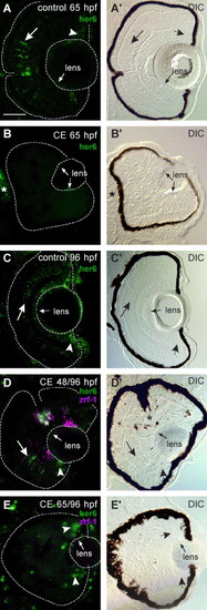
Lack of her6/hes1 expression reflects inhibition of Notch signaling. Cryosections of control and γ-secretase-inhibited (CE) embryos labeled for her6/hes1 expression (FITC—green) by in situ hybridization. DIC images indicate the location of her6/hes1 expression in the retina (outlined) in the paired panels to the left. (A, A′) At 65 hpf, her6/hes1 expression in the inl (large arrow) and germinal zone (arrowhead) of the retina and in the lens epithelium (small arrow) in control embryos. (B, B′) her6/hes1 expression in the lens epithelium (small arrows) and brain (*) of CE-treated embryos (N = 15 embryos). (C, C′) At 96 hpf, her6/hes1 is expressed in the inl, often in radial streaks (large arrow), and in the germinal zone (arrowhead) of the retina and in the lens epithelium (small arrow) of control embryos (N = 15 embryos). A faint reaction product is also seen in the gcl, but this may be localized to the endfeet of Müller glia—a similar pattern of gfap mRNA expression is observed in retinas at this stage (R.L.B., unpublished observations). (D, D2) In embryos treated with CE up to 48 hpf and then allowed to develop in drug-free embryo media up to 96 hpf, her6/hes1 is expressed in the inl (large arrow), germinal zone of the retina (arrowhead), the optic nerve head (*; astrocytes) and in the lens epithelium (small arrow). Co-labeling with zrf-1/GFAP (Cy-3-pseudo-colored magenta) reveals a correlation between the location of zrf-1/GFAP and her6/hes1 expression in the inl (N = 15 embryos). (E, E2) In embryos treated with CE up to 65 hpf and then allowed to develop in drug-free embryo media up to 96 hpf, her6/hes1 is expressed only in the retinal germinal zone (arrowhead) and lens epithelium (small arrows) and no zrf-1/GFAP immunoreactivity is detected in the retina (N = 15 embryos). Scale bar = 50 μm (A–E). |
| Genes: | |
|---|---|
| Fish: | |
| Condition: | |
| Anatomical Terms: | |
| Stage Range: | Pec-fin to Day 4 |
Reprinted from Developmental Biology, 278(2), Bernardos, R.L., Lentz, S.I., Wolfe, M.S., and Raymond, P.A., Notch-Delta signaling is required for spatial patterning and Muller glia differentiation in the zebrafish retina, 381-395, Copyright (2005) with permission from Elsevier. Full text @ Dev. Biol.