FIGURE SUMMARY
Title

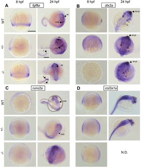
Fgf8a mutation affects craniofacial development and skeletal gene expression in zebrafish larvae

Authors
Gebuijs, I.G.E., Raterman, S.T., Metz, J.R., Swanenberg, L., Zethof, J., Van den Bos, R., Carels, C.E.L., Wagener, F.A.D.T.G., Von den Hoff, J.W.
Source
Full text @ Biol. Open

Representative examples of a 5 dpf wild-type (A), fgf8ati282 heterozygous (B) and -homozygous (C) larvae. Larvae were stained for cartilage (blue) and bone (red). Note the difference in head size; scale bar: 200 µm. Also note the absence of mineralization, such as ceratobranchial 5 and the parasphenoid bone in the hetero- and homozygote (arrowheads), and the presence of teeth in the homozygote (arrows). Note the considerably smaller Meckel's cartilage in hetero- and homozygotes (double-headed arrows).

ZFIN is incorporating published figure images and captions as part of an ongoing project. Figures from some publications have not yet been curated, or are not available for display because of copyright restrictions.

ZFIN is incorporating published figure images and captions as part of an ongoing project. Figures from some publications have not yet been curated, or are not available for display because of copyright restrictions.

ZFIN is incorporating published figure images and captions as part of an ongoing project. Figures from some publications have not yet been curated, or are not available for display because of copyright restrictions.

Early spatiotemporal gene expression inacemutant development. Wholemount in situ hybridization in 8 hpf and 24 hpf wild-type, heterozygous and homozygous embryos shows patterns of early gene expression of (A) fgf8a, (B) dlx2a, (C) runx2a and (D) col2a1a. mhb, mid-hindbrain boundary; dd, dorsal diencephalon; t, tail end; s, somites; pt, prethalamus; tel, telencephalon; fb, forebrain; hb, hindbrain. N.D.: no data were obtained. Scale bar: 200 µm, in insert: 50 µm.

5 dpf zebrafish larva (wild type) stained for cartilage (blue) and bone (red). A total of nine parameters for cartilage were assessed as shown in panels A (ventral view) and B (lateral view): (1) total head length, (2) length ceratohyal to anterior end of the head, (3) length ceratohyal to posterior end of the head, (4) width at Meckel's cartilage and palatoquadrate joint, (5) width at ceratohyal and palatoquadrate joint, (6) length of superior ceratohyal, (7) length of inferior ceratohyal, (8) length of ethmoid plate, (9) lateral length of Meckel's cartilage. (C) The nine bone parameters scored for presence or absence (ventral view): (1) parasphenoid, (2) cleithrum, (3) notochordal sheath, (4) otoliths (four in total), (5) ceratobranchial 5, (6) teeth (on ceratobranchial 5), (7) opercles, (8) branchiostegal ray 1, (9) entopterygoid bone.

Acknowledgments
This image is the copyrighted work of the attributed author or publisher, and ZFIN has permission only to display this image to its users. Additional permissions should be obtained from the applicable author or publisher of the image. Full text @ Biol. Open