
- Title
-
Transcriptome analysis of pancreatic cells across distant species highlights novel important regulator genes
- Authors
- Tarifeño-Saldivia, E., Lavergne, A., Bernard, A., Padamata, K., Bergemann, D., Voz, M.L., Manfroid, I., Peers, B.
- Source
- Full text @ BMC Biol.

Expression of genes in endocrine cells of the dorsal pancreatic bud. Whole-mount in situ hybridization on zebrafish embryos showing endocrine pancreatic expression of some new zebrafish endocrine markers (n > 15). Genes with conserved endocrine-enriched expression in zebrafish, human, and mouse (ZHM), or endocrine-enriched in zebrafish and mouse (ZH), or in zebrafish and human (HM) are indicated. Z: Gene endocrine enriched only in zebrafish. N-orth: Endocrine enriched zebrafish gene with no obvious human or mouse ortholog. Arrowheads indicate the location of the dorsal pancreatic bud containing embryonic endocrine cells and insets at the top-right display higher magnification view of the pancreatic bud |

Expression of ppdpfb and pcsk2 genes in zebrafish beta cells. Co-labeling by fluorescent in situ hybridization (FISH) of ppdpfb and pcsk2 with insulin, glucagon, and somatostatin. a–c ppdpfb is mainly expressed by beta cells (a, arrows) and in few alpha cells (b, arrows). No expression of ppdpfb was observed in delta cells (c). Beta cells specifically expressed pcsk2 (d, arrows) while no expression was detected in alpha or delta cells (e and f). (Analyzed embryos > 10) EXPRESSION / LABELING:
|
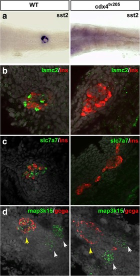
Delta cell differentiation is disrupted in the cdx4 mutant embryos. Analysis of wild-type and cdx4 tv205–/– mutant embryos at 48 hpf by WHISH (a) and FISH (b–d) using delta cell markers (sst2, lamc2, slc7a7, and map3k15) as well as gcga and insulin. cdx4 tv205 mutants display a loss of sst2 (a), lamc2 (b), and slc7a7 (c) pancreatic expression and an increase of beta cells. Map3k15 expression is strongly reduced in the pancreatic islet (yellow arrows) while not affected in the presumptive pronephric glomeruli (white arrows) (d). No obvious effect is observed on gcga expression. Nuclei are stained with DAPI (grey staining). (Analyzed embryos > 10) |

ZFIN is incorporating published figure images and captions as part of an ongoing project. Figures from some publications have not yet been curated, or are not available for display because of copyright restrictions. |
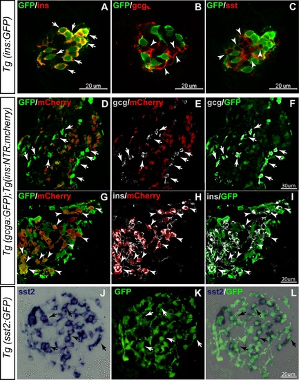
Expression of the transgenes Tg(ins:GFP), Tg(gcga:GFP), Tg(sst2:GFP), and Tg(ins:NTR:mCherry) in the endocrine pancreatic cell types. (A–C) Whole mount immunostaining of 3 dpf transgenic larvae Tg(ins:GFP) demonstrating the selective expression of GFP in beta cells. (D–I) Immunostaining of pancreas sections from adult transgenic fish Tg(gcga:GFP/ins:NTR:mCherry). Glucagon+ cells (see arrows in D–F) express high level of GFP and are not labelled by mCherry, while many insulin expressing cells are labelled by mCherry and by GFP (at slightly lower levels) (see arrowheads in G–I). These data reveal a leaking expression of the gcga:GFP transgene in beta cells. (J–L) ISH performed on pancreas section of adult Tg(sst2:gfp) with sst2 probe followed by immunofluorescence using the GFP antibody; the expression of endogenous sst2 gene co-localize with GFP staining confirming the specific expression of the transgene in delta cells [28]. EXPRESSION / LABELING:
|
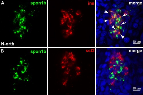
Expression of spon1b gene in beta pancreatic cells of zebrafish embryos. Co-labeling by FISH of spon1b with insulin (ins) (A, arrows show colocalization), while no expression was detected in delta cells (B) (n > 10). ins: insulin, sst2: somatostatin 2, N-orth: Endocrine enriched zebrafish gene with no described ortholog in human and/or mouse. EXPRESSION / LABELING:
|

Expression of pnoca and scinlb genes in alpha pancreatic cells of zebrafish embryos. Co-labeling by in situ hybridization at 30 hpf of new discovered genes with cell type-specific markers for alpha and beta cells (n > 10). A and B. pnoca is expressed in alpha cells (A, arrows) while no expression was detected in beta cells (B). scinlb was detected specifically in alpha cells (C, arrows) but not detected in beta cells (D). gcga: glucagon a, ins: insulin, Z: no endocrine gene with no conserved expression, N-orth: Endocrine-enriched zebrafish gene with no described ortholog in human and/or mouse. EXPRESSION / LABELING:
|

Validation of the selective expression of some genes in zebrafish delta cells. cdx1b, cdx4, lamc2, and map3k15 are specifically expressed in delta cells at 24 hpf (A, C, E, G, arrows show colocalization with delta cell-specific markers, somatostatin 2; n > 10). No expression was detected for none of the genes in beta cells (B, D, F, H). ins: insulin, sst2: somatostatin 2, ZHM: Gene expression conserved in zebrafish, human and mouse, ZM: Gene expression conserved in zebrafish and mouse, Z: Endocrine gene with no conserved expression, N-orth: Endocrine-enriched zebrafish gene with no described ortholog in human and/or mouse. EXPRESSION / LABELING:
|
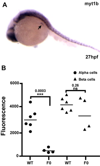
Expression and function of myt1b in zebrafish pancreas. A: Whole-mount ISH showing expression pattern of myt1b in zebrafish embryos at 27 hpf. High expression is detected in the dorsal pancreatic bud (indicated by the arrow) and in the central nervous system. B: Quantification of glucagon and insulin expression at 48 hpf in wild-type (non-injected) embryos and in embryos injected with the 4 CRISPR myt1a/b guide RNA and Cas9. Graph B shows the volume of all gcga+ cells and all ins+ cells measured in each embryo by the imaging software Imaris (see Methods) (each point is the volume measured in one embryo). This quantification indicates a statistically significant reduction of the volume of alpha cell mass while beta cell mass is not drastically affected in the injected (F0) embryos (results of one experiment). |