
- Title
-
Global analysis of the haematopoietic and endothelial transcriptome during zebrafish development
- Authors
- Cannon, J.E., Place, E.S., Eve, A.M., Bradshaw, C.R., Sesay, A., Morrell, N.W., and Smith, J.C.
- Source
- Full text @ Mech. Dev.

Genes with blood and vascular in situ hybridisation expression pattern in 24–28hpf embryos. All embryos are lateral views with anterior to left. Scale bars indicate 500 μm. High powered views of tail region of these embryos are shown in Supplementary Fig. 4. |
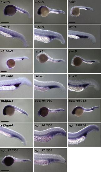
Genes with blood in situ hybridisation expression pattern in 24–28hpf embryos. All embryos are lateral views with anterior to left. Scale bars indicate 500 μm. |
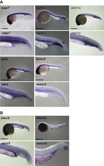
Remaining in situ hybridisation expression patterns in 24–28hpf embryos. (A) Vascular expression (B) Other. All embryos are lateral views with anterior to left. Scale bars indicate 500 μm. EXPRESSION / LABELING:
|

Tmem88a and trim2a morphants have reduced erythrocytes and myeloid cells as shown by O-dianisidine and peroxidase staining respectively at 48 h post fertilisation. In control embryos erythrocytes are present in axial vessels and returning to the heart across the yolk. Myeloid cells are found across the yolk and randomly around the remaining parts of the embryos. There is loss of erythrocytes and myeloid cells in both trim2a and tmem88a morphants (translation and splice blocking morpholinos) without any defect in vascular development. Panel row 1 shows representative brightfield images, row 2 epifluorescent images of Tg(fli1a:egfp) embryos, rows 3 and 4 show embryos post O-dianisidine staining and row 5 show embryos post peroxidase staining. All images are lateral views with anterior to left with the exception of row 4 which are ventral views. PHENOTYPE:
|
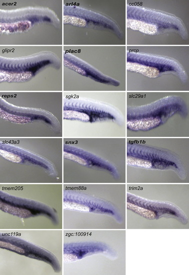
High powered tail view of embryos with blood and vascular cell in situ hybridisation expression patterns in 24–28 hpf embryos. All embryos are lateral views with anterior to left. |
Reprinted from Mechanisms of Development, 130(2-3), Cannon, J.E., Place, E.S., Eve, A.M., Bradshaw, C.R., Sesay, A., Morrell, N.W., and Smith, J.C., Global analysis of the haematopoietic and endothelial transcriptome during zebrafish development, 122-131, Copyright (2013) with permission from Elsevier. Full text @ Mech. Dev.