
- Title
-
Tracking gene expression during zebrafish osteoblast differentiation
- Authors
- Li, N., Felber, K., Elks, P., Croucher, P., and Roehl, H.H.
- Source
- Full text @ Dev. Dyn.

A-Y: Osteoblast gene expression: side views. Five stages of development are shown highlighting the development of the two related cranial bones, the third brachiostegal ray and the opercle, as well as the cleithrum, which is part of the shoulder. In addition, the parasphenoid and fifth ceratobranchial are labeled in F and H. The double arrow in L marks the distal edge of the second arch for reference. All views are of the left side of the head, focusing on the plane of the opercle, with the eye and yolk visible to either side (labeled in A). bs, brachiostegal ray; cb5, ceratobranchial arch 5 (including teeth); ch, ceratohyal; cl, cleithrum; de, dentary; en, entopterygoid; hm, hyomandibular; mx, maxilla; op, opercle; ps, parasphenoid. A high resolution image of this figure is available online. Scale bar = 100 μM in Y. |

A-Z: Osteoblast gene expression: dorsal and ventral views. Six stages of development are shown in dorsal views at 36 and 48 hours postfertilization (hpf; A,B,G,H,M,N,S,T,Y,Z), and in ventral views at 63, 72, 96, and 120 hpf (C-F,I-L,O-R,U-X,AA-DD). A high resolution image of this figure is available online. See Figure 1 for abbreviations. Scale bar = 200 μM in DD. |

Atypical osteoblast gene expression: side views. Five stages shown as described in Figure 1. See Figure 1 for abbreviations and scale bar. A high resolution image of this figure is available online. |
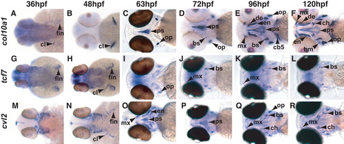
A-R: Atypical osteoblast gene expression: dorsal and ventral views. Six stages of development are shown highlighting the development the cleithrum in dorsal views at 36 and 48 hpf (A,B,G,H,M,N), and the development of other bones in ventral views at 63, 72, 96, and 120 hpf (C-F,I-L,O-R). See Figure 1 for abbreviations and Figure 2 for scale bar sizing. A high resolution image of this figure is available online. |
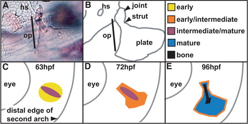
Model for osteoblast differentiation during formation of the opercle. A: Alizarin Red staining of the fan-shaped opercle (op) at 144 hours postfertilization (hpf) showing its connection to the hyosymplectic (hs) cartilage proximally. B: Camera lucida drawing of A showing the joint, strut, and plate of the opercle. C-E: See text for explanation. |