
- Title
-
Distinct requirements of wls, wnt9a, wnt5b and gpc4 in regulating chondrocyte maturation and timing of endochondral ossification
- Authors
- Ling, I.T., Rochard, L., Liao, E.C.
- Source
- Full text @ Dev. Biol.

Overlapping domains of gene expression ofwls,wnt9a,wnt5bandgpc4in ventral craniofacial cartilages. (A-D) 10X Whole mount RNA in situ hybridization with ventral views with 40X focused on Meckel's cartilage (A′-D′). At 96 hpf, wls (A) is expressed in the surrounding tissue including Meckel's cartilage and co-expressed with wnt9a (B) and gpc4 (D) in the posterior ceratobranchials (light blue). In contrast, wnt5b appeared to be expressed in the chondrocytes of the ceratobranchials and the blood vessels surrounding them (C). wls, wnt5b and gpc4 co-localized in the surrounding mesenchyme around the jaw joint (arrowheads in A′-D′ and green in diagram E). wnt5b is expressed in the oral epithelium with wls. There is discrete expression of wnt9a in the anterior midline (purple) adjacent to the intermandibulare anterior muscle. Annotation: Meckel's cartilage (mc), palatoquadrate (pq), hyosymplectic (hs), interhyal (ih), ceratobranchial (cb), ceratohyal (ch), basihyal (bh). Scale=50 µm. EXPRESSION / LABELING:
|
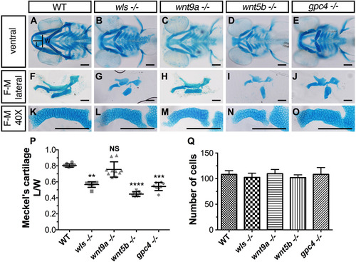
Cartilage morphogenetic defects inwls,wnt9a,wnt5bandgpc4mutants. (A-E) Ventral view of whole-mount 96 hpf Alcian blue stain of wls, wnt9a, wnt5b and gpc4. (F-J) Dissected flat-mounted lateral image of ventral cartilages. (K-O) 40X image of the Meckel's cartilage lateral view. Length (L) calculated from the midline tip of the Meckel's cartilage to the middle of the imaginary line (width (W)) between the retroarticular processes. (P) Meckel's cartilage length/width ratio measured from a clutch of imaged embryos (**p<0.01, ***p<0.001, ****p<0.0001; Kruskal-Wallis test with Dunn's multiple comparison test. Significance level at p<0.05). (Q) Number of cells from one-half of Meckel's cartilage was counted from figures K-O. No statistically significant difference in cell number across wls, wnt9a, wnt5b and gpc4 (p=0.1151, NS; One-way ANOVA). Scale =50 µm.. PHENOTYPE:
|
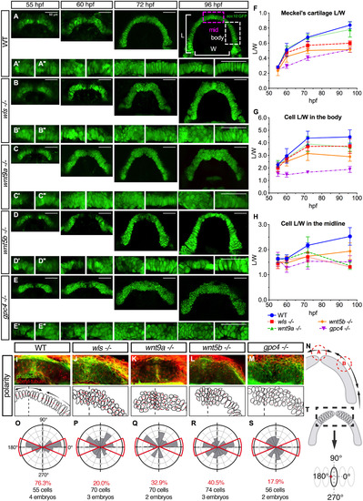
: Distinct cellular and polarity differences between midzone and body of Meckel's cartilage in wls, wnt9a, wnt5b and gpc4 mutants. (A-E) Wnt gene were crossed to Tg(sox10:GFP) transgenic line and homozygote embryos were imaged at difference time points starting at 55 hpf when convergence of the Meckel's cartilage occurs until 96 hpf when the arch is complete. Defects evident from 60 hpf especially in gpc4 -/- and by 72 hpf, obvious midline defect was seen in wls, wnt9a and gpc4 and body defects apparent in gpc4. wnt5b exhibited a smaller cartilage size throughout morphogenesis. (A; 96 hpf) Graphical representation of the L/W measurements from Meckel's cartilage. (A’) 20X image of Meckel's cartilage in the midline and (A′’) 20X image of the body of Meckel's. (F-H) Individual cell length/width ratio were measured and plotted over time from 2 distinct parts of the Meckel's; midzone and body as marked in the illustration. (I-M) 72 hpf WT and homozygote embryos were stained for actin with phalloidin (green) to delineate cell membranes and acetylated-tubulin (red) to reveal microtubule organizing center (MTOC). (N) Polarity pattern for Meckel's cartilage at 72 hpf showing 2 inflection points; point A where the hemi-Meckel's cartilage meet in the midline and point B within the body of each hemi-Meckel's cartilage. (O-S) Rose plots of cell polarity as indicated by MTOC measured from cells within the midzone portion of Meckel's cartilage. (T; black dotted box). Scale=50 µm. PHENOTYPE:
|

Bone defects inwls,wnt9a,wnt5bandgpc4mutants. (A-J) Maximum intensity projections of live confocal imaging stacks of 4 dpf (A-E) and 8 dpf (F-J) Wnt homozygotes in Tg(sox10:GFP) background stained with alizarin red S. (K-O) 40X confocal maximum intensity projections of alizarin red stain focusing on the dentary bone (de) and mentomeckelian (mm). The mentomeckelian is seen as a flatter bone attached to the distal end of the dentary that is thicker. A clear boundary is observed between the mm and de. wls mutants lacks the dentary or mentomeckelian. (P-T) Confocal maximum intensity projections of osteocalcin transgene expression in Wnt mutants in a Tg(sox10:mCherry) background. (U-Y) Confocal maximum intensity projections of osx transgene expression in Wnt mutants in a Tg(sox10:GFP) background. (Z) Graphical map of neural-crest cell (NCC)-derived bone (red; intramembranous ossification and orange; endochondral ossification) and mesoderm derived bone (blue). Mentomeckelian (mm), Maxilla (mx), Dentary (de), retroarticular (ra), quadrate (q), ceratohyal (ch), branchiostegal rays (bsr), opercle (op), ceratobranchial 5 (cb5), cleithrum (cl). (AA) Percentage of mutant with bony phenotype of different bones in the craniofacial region where 100% indicates that all mutant embryos do not have that particular bone. Scale=50 µm. |

Joint and muscle defects inwls,wnt9a,wnt5b, andgpc4mutants. (A-E) Alcian blue stained ventral cartilage lateral whole-mounts of 8 dpf embryos and 40X zoomed image in A′-E′ of the jaw joint (jj; left) and hinge joint (hj; right). Meckel's cartilage (mc), pterygoid process (ptp), palatoquadrate (pq), hyosymplectic (hs), and ceratohyal (ch). Jaw joint defects evident in wls and gpc4 mutant whilst wnt9a exhibited a slightly malformed retroarticular process. wls displayed a clump of cells within the hinge-joint compared to the neatly stacked layered chondrocytes in wild-type. In contrast, wnt9a, wnt5b and gpc4 were only a single cell layer in thickness. (F-J) Maximum intensity projections of 20X zoom confocal stacks showing muscle stain with phalloidin (purple) and anti-GFP (green) for cartilage elements. Muscle length defects apparent in all Wnt mutants with gpc4 displaying disorganized intermandibularis posterior (imp) invading the gaps in Meckel's cartilage. Adductor mandibulae (am), intermandibularis anterior (ima), intermandibularis posterior (imp), hyohyoideus (hh), interhyoideus (ih), sternohyoides (sh) F′-J’’ Single channel phalloidin flourescence. (K) Graphical representation of retroaritcular measurement. (L) Measured length of retroarticular process and (M) cell count per interhyal in WT and Wnt mutants. (*p<0.01***p<0.001, ****p<0.0001; Kruskal-Wallis test with Dunn's multiple comparison test. Significance level at p<0.05). Scale =50 µm.. |
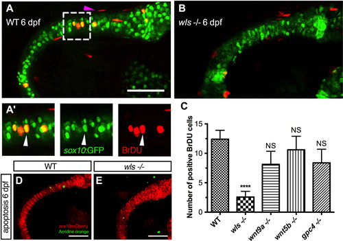
Chondrocyte proliferation defect inwlsmutant precedes the differentiation defect. (A, B) Representative BrdU assay confocal stacked image in WT 6 dpf (A) and wls -/- (B) embryo with zoomed views of WT BrdU (red) colocalizing with chondrocytes (A’) in the Meckel's cartilage (green). Arrowhead points to a representative chondrocyte that expressed both sox10 and BrdU and used for quantification. (C) Quantification of BrdU positive cells in Wnt mutants. Proliferation significantly differed in wls -/- compared to WT (****p<0.001; Kruskal-Wallis test and Dunn's multiple comparison test, NS = not significant). (D, E) No difference in chondrocyte apoptosis as showed with Acridine orange live staining at 6 dpf in wls -/-. Scale=50 µm. PHENOTYPE:
|
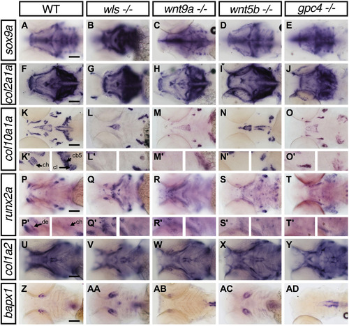
Lower jaw gene expression pattern of chondrocyte and bone markers inwls,wnt9a,wnt5bandgpc4mutants. Whole-mount RNA in situ hybridization of chondrocyte differentiation markers; sox9a (A-E) and chondrocyte matrix; col2a1 (F-J). Hypertrophic chondrocyte marker; col10a1a (K-O), Bone and tendon marker; col1a2 (P-T), osteoblastic progenitor marker; runx2a (U-Y) and joint marker; bapx1 (Z-AD). (K′-L′) 40X image focused on ceratohyal (ch; left) and both cleithrum and ceratobranchial 5 (cl, cb; right). (P′-T′) 40X images focused on dentary (de; left) and ceratohyal (right). Apparent increase in sox9a, runx2a expression pattern in wls -/- with concomitant loss of col10a1a. Black arrowhead points to the position of Meckel's cartilage. Scale=50 µm.. |
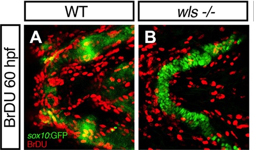
Supplementary material: Supplementary Fig 1. No difference in cell proliferation between WT and wls mutants at 60 hpf. Maximum intensity projections of confocal Z-stacks for BrdU assay in WT larvae Tg(sox10:GFP) at 60 hpf (A) and wls -/- (B). PHENOTYPE:
|
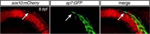
Supplementary material: Supplementary Fig 2. Osterix-expressing cells in 8 dpf WT embryos. 40X Confocal maximum intensity projection images of 8 dpf Meckel's cartilage. Osterix expressing cells are observed at the position of the dentary adjacent to the body of Meckel's above the perichondrium. A single osterix-expression cell (arrow) is observed at the level of the perichondrium indicating the differentiation of perichondral cells to osteoblast to form the future mentomeckelian perichondral bone. EXPRESSION / LABELING:
|
Reprinted from Developmental Biology, 421(2), Ling, I.T., Rochard, L., Liao, E.C., Distinct requirements of wls, wnt9a, wnt5b and gpc4 in regulating chondrocyte maturation and timing of endochondral ossification, 219-232, Copyright (2017) with permission from Elsevier. Full text @ Dev. Biol.