
- Title
-
Neucrin, a novel secreted antagonist of canonical Wnt signaling, plays roles in developing neural tissues in zebrafish
- Authors
- Miyake, A., Nihno, S., Murakoshi, Y., Satsuka, A., Nakayama, Y., and Itoh, N.
- Source
- Full text @ Mech. Dev.

Expression of neucrin in zebrafish embryos. Expression pattern of neucrin in zebrafish embryos at the indicated stages detected by whole-mount in situ hybridization. (A-D, F) Dorsal view with anterior to the top. (E, G-K) Lateral view with anterior to the left and dorsal to the top. Long arrows and arrowheads indicate the spinal cord and somite, respectively. A short arrow in E indicates tightly clustered cell groups in the ventrorostral forebrain. di, diencephalon; hb, hindbrain; mb, midbrain; te, telencephalon. Scale bar: 100 μm in A-H, K; 25 μm in I, J. EXPRESSION / LABELING:
|
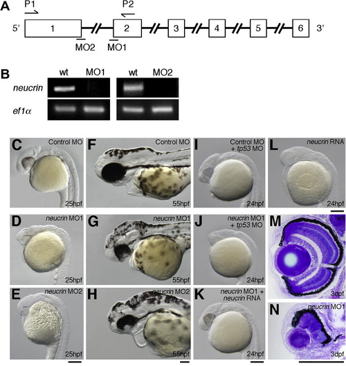
Inhibition of Neucrin functions in zebrafish embryos. (A) The coding region of neucrin is divided by five introns. Open boxes and black lines indicate exons and introns, respectively. MO indicates the target position of neucrin MO. (B) neucrin cDNA was amplified from wild-type or neucrin MO-injected embryonic cDNA by RT-PCR using forward and reverse primers, the positions of which are indicated by arrows (A). ef1α cDNA was also amplified as a control. (C-H) Lateral view of control MO-injected (C, F), neucrin MO1-injected (D, G) and neucrin MO2-injected (E, H) embryos at the indicated stages are shown. MOs were injected into four central blastomeres of 16-cell embryos. (I, J) Lateral view of tp53 MO- and control MO-injected (I), and tp53 MO- and neucrin MO1-injected (J) embryos at the indicated stages are shown. tp53 MO was injected into 2- to 4-cell embryos. neucrin MO1 and control MO were injected into four central blastomeres of 16-cell embryos. (K, L) Lateral view of neucrin RNA- and neucrin MO1-injected (K), and neucrin RNA-injected (L) embryos at the indicated stages are shown. neucrin MO1 was injected into four central blastomeres of 16-cell embryos. RNA was injected into 2- to 4-cell embryos. (M, N) Transverse eye sections of wild-type (M) and neucrin MO1-injected (N) embryos at 3 dpf. Scale bar: 100 μm. PHENOTYPE:
|

Effect of neucrin on the expression of Wnt/β-catenin target genes. (A-D) The expression of axin2 (A, B) and cyclin D1 (C, D) in wild-type (A, C) and neucrin MO1-injected (B, D) embryos at 24 hpf. Lateral view with anterior to the left. (E, F) The expression of axin2 in wild-type (E) and neucrin RNA-injected (F) embryos at 24 hpf. Dorsal view with anterior to the left. (G) qRT-PCR analysis on mRNA generated from wild-type embryos and embryos injected with neucrin MO1-injected and neucrin RNA-injected embryos. Results are the means ± S.D. (n = 3). The statistical significance of differences in mean values was assessed with the Student’s t-test. Asterisks indicate statistical significance compared with the wild type (*P < 0.05; **P < 0.01). (H-J) Dorsal view of sp5l expression in a wild-type embryo in which wild-type cells (H) or neucrin-overexpressing cells (I, J) were transplanted. Transplanted cells are labeled in brown. neucrin-overexpressing donor cells transplanted into the axis (I) and non-axial mesoderm (J) did not express sp5l (white arrows) and suppressed sp5l expression in the surrounding host regions (white arrowheads) at 9 hpf. Scale bar: 100 μm. EXPRESSION / LABELING:
|
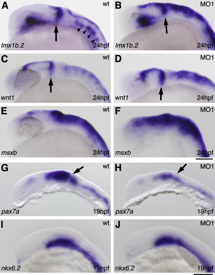
Effect of neucrin on dorsal CNS development. The expression of lmx1b.2 (A, B), wnt1 (C, D), msxb (E, F), pax7a (G, H) and nkx6.2 (I, J) in wild-type (A, C, E, G, I) and neucrin MO1-injected (B, D, F, H, J) embryos at 24 hpf. Lateral view with anterior to the left. Arrows indicate the MHB. Arrowheads indicate serotonergic neurons. Scale bar: 50 μm. EXPRESSION / LABELING:
|
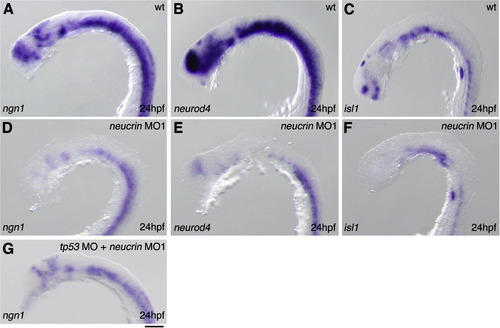
Effect of neucrin on neuronal differentiation. (A-G) The expression of ngn1 (A, D, G), neurod4 (B, E) and isl1 (C, F) in wild-type (A, B, C), neucrin MO1-injected (D, E, F), and tp53 MO- and neucrin MO1-injected (G) embryos at 24 hpf. Lateral view with anterior to the left. Scale bar: 50 μm. EXPRESSION / LABELING:
|
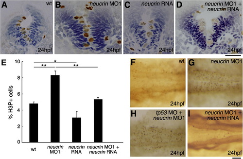
Comparison of cell proliferation and death in neucrin MO1-injected and neucrin RNA-injected embryos. (A-E) Wild-type embryos (A) and embryos injected with neucrin MO1 (B), neucrin RNA (C), or neucrin MO1 and neucrin RNA (D) were stained using an anti-phosphorylated Histone H3 antibody. (A-D) Panels show representative transverse sections of the hindbrain at 24 hpf. Brown and blue cells indicate the H3P-positive cells and H3P-negative cells, respectively. (E) The percentage of proliferating cells labeled with anti-H3P antibody in the hindbrain of wild-type embryos and embryos injected with neucrin MO1 or neucrin RNA or co-injected with neucrin MO1 and neucrin RNA. Results are the means ± S.D. for three embryos. The statistical significance of differences in mean values was assessed with the Student’s t-test. Asterisks indicate statistical significance compared with the wild type (*P < 0.05; **P < 0.01). (F-I) At 24 hpf, apoptotic cells in the hindbrain of wild-type embryos (F) and embryos injected with neucrin MO1 (G), neucrin MO1 and p53 MO (H), or neucrin MO1 and neucrin RNA (I) were marked via TUNEL labeling. Dorsal view with anterior to the left. Scale bar: 50 μm. PHENOTYPE:
|
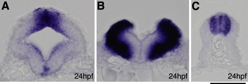
Expression of neucrin in zebrafish embryos at 24 hpf. (A) Transverse section through the midbrain. (B) Transverse section through the hindbrain. (C) Transverse section through the spinal cord. Scale bar: 50 μm. |

Morphology in the embryos injected with neucrin MOs at the 2-cell stage. Dorsal view of wild-type (A), neucrin MO1-injected (B), neucrin MO2-injected (C), or neucrin RNA- and neucrin MO1-injected (D) embryos at the indicated stages are shown. Scale bar: 100 μm. PHENOTYPE:
|

Morphology in the embryos injected with neucrin MOs at the 16-cell stage. Dorsal view of control MO-injected (A), neucrin MO1-injected (B), neucrin MO2-injected (C), tp53 MO- and control MO-injected (D), tp53 MO- and neucrin MO1-injected (E), neucrin MO1- and neucrin RNA-injected (F), or neucrin RNA-injected (G) embryos at 24 hpf are shown. Scale bar: 100 μm. PHENOTYPE:
|

Effect of neucrin on the expression of targeted genes of Wnt/β-catenin signaling. (A–D) The expression of sp5l in wild-type (A), wnt8a RNA-injected (B), neucrin RNA-injected (C), and wnt8a RNA- and neucrin RNA-injected (D) embryos at 8 hpf. Dorsal view with anterior to the top. (E and F) The expression of otx2 in wild-type (E) and neucrin RNA-injected (F) embryos at 8 hpf. Lateral view with anterior to the left. Scale bar: 100 μm. |
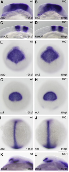
Effect of neucrin on AP patterning and non-neuronal differentiation. (A–D) The expression of otx2 (A and B) and krox20 (C and D) in wild-type (A and C) and neucrin MO1-injected (B and D) embryos at 19 hpf. Lateral view with anterior to the left. (E–H) The expression of otx2 (E and F) and rx3 (G and H) in wild-type (E and G) and neucrin MO1-injected (F and H) embryos at 10 hpf. Dorsal view with anterior to the top. (I and J) The expression of ntla in wild-type (I) and neucrin MO1-injected (J) embryos at 11 hpf. Dorsal view with anterior to the top. (K and L) The expression of dlx2a in wild-type (K) and neucrin MO1-injected (L) embryos at 16 hpf. Lateral view with anterior to the left. Scale bar: 100 μm. |

Results of RNA rescue experiments. The expression of lmx1b.2 (A), ngn1 (B) and isl1 (C) in neucrin RNA- and neucrin MO1-injected embryos at 24 hpf. Lateral view with anterior to the left. Scale bar: 50 μm. |
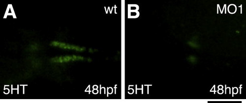
Effect of neucrin on the differentiation of serotonergic neurons. Dorsal views of wild-type (A) and neucrin MO1-injected (B) embryos, labeled to show 5HT immunoreactivity. Scale bar: 100 μm. |
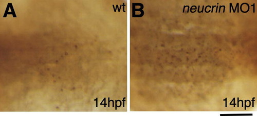
Apoptosis in the hindbrain of neucrin MO1-injected embryos at 14 hpf. (A and B) At 14 hpf, apoptotic cells in the hindbrain of the wild-type (A) and neucrin MO1-injected (B) embryos were marked via TUNEL labeling. Dorsal view with anterior to the left. Scale bar: 100 μm. |
Reprinted from Mechanisms of Development, 128(11-12), Miyake, A., Nihno, S., Murakoshi, Y., Satsuka, A., Nakayama, Y., and Itoh, N., Neucrin, a novel secreted antagonist of canonical Wnt signaling, plays roles in developing neural tissues in zebrafish, 577-590, Copyright (2012) with permission from Elsevier. Full text @ Mech. Dev.