
Fig. 3
- ID
- ZDB-FIG-170321-25
- Publication
- Liu et al., 2017 - Stat3/Cdc25a-dependent cell proliferation promotes embryonic axis extension during zebrafish gastrulation
- Other Figures
- All Figure Page
- Back to All Figure Page
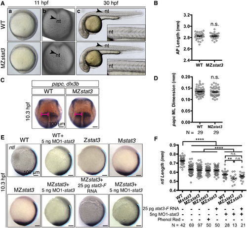
MZstat3 mutants exhibit transient and mild extension defects in axial mesoderm during gastrulation. (A) Live images of WT and MZstat3 embryos shown in lateral (a, c) and dorsal view (b). The insets in c show the part of notochord above yolk extension in 30 hpf embryos. Arrowhead, nt, notochord. (B) Morphometric analysis of AP axis extension of 30 hpf embryos shown in A(c). (C) papc in presomitic mesoderm and dlx3b marking neuroectoderm boundary in 1-somite stage WT and MZstat3 embryos (dorsal view). (D) Measurement of ML width of papc expression domain (pink in C). (E) Expression of ntl in notochord and tail in 1-somite stage WT, Zstat3, Mstat3, MZstat3, and MZstat3 embryos overexpressing Stat3-F, as well as WT and MZstat3 embryos injected with 5 ng MO1-stat3 (lateral view). Phenol red was used as injection control. (F) Measurement of notochord length in embryos in E (blue lines in E). ****p<0.0001, n.s. = non-significant, error bars = SEM. See also S3 Fig. |
| Genes: | |
|---|---|
| Fish: | |
| Knockdown Reagent: | |
| Anatomical Terms: | |
| Stage: | 1-4 somites |
| Fish: | |
|---|---|
| Knockdown Reagent: | |
| Observed In: | |
| Stage: | 1-4 somites |