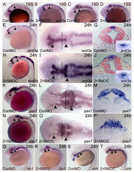
Knockdown of zic2a and zic5 disrupts midbrain morphology, but not DV pattern. Embryos were injected with conMO (8 ng), or 2MOC and 5MOC combined (1 ng each), and stained for DV marker expression by ISH. (A-D) Marker expression in 19-somite embryos. wnt3a marks dorsal midline and adjacent cells in midbrains of conMO-injected embryos (A) and in zic morphants (B). pax7 marks alar midbrain in controls (C) and morphants (D). (E-T) Marker expression in 24-hpf embryos. wnt3a is expressed in dorsal tectum of conMO-injected embryos (E-G). (H-J) wnt3a domain in zic morphants. (K-M) pax7 in the alar tectum of controls. (N-P) pax7 in zic morphants is reduced, but correctly positioned. (Q,R) hlx1 marks basal plate midbrain of controls (Q) and zic morphants (R). (S,T) shh in ventral midbrain of controls and zic morphants. A-D,E,H,K,N,Q-T are lateral views, anterior to the left; F,I,L,O are dorsal views of embryos at left; G,J,M,P are midbrain cross-sections at positions indicated by lines in images to left. Arrowheads indicate the midbrain.
|
