FIGURE SUMMARY
Title

Rab5c-mediated endocytic trafficking regulates hematopoietic stem and progenitor cell development via Notch and AKT signaling

Authors
Heng, J., Lv, P., Zhang, Y., Cheng, X., Wang, L., Ma, D., Liu, F.
Source
Full text @ PLoS Biol.

Endocytic trafficking regulator Rab5c is highly expressed in embryonic hematopoiesis region.

(A) Flowchart of FACS and RNA-seq analysis of the different types of cells in the trunk region from Tg(kdrl:mCherry/runx1:en-GFP) transgenic zebrafish embryos at about 28 hpf. (B) GO analysis of the sequencing data shows that up-regulated transcripts in runx1+ HE are enriched in the endosome and endocytic trafficking related terms. (C) RPKM values of Rab5 family genes in EC and HE. (D) Expression pattern of rab5c mRNA examined by WISH and double FISH. Scale bar, 200 μm. The P values in this figure were calculated by Student t test. The underlying data in this figure can be found in S1 Data. CA, caudal vein; DA, dorsal aorta; EC, endothelial cell; FACS, fluorescence-activated cell sorting; FISH, fluorescence in situ hybridization; GO, gene ontology; HE, hemogenic endothelium; hpf, hours post fertilization; RNA-seq, RNA sequencing; RPKM, reads per kilobase of exon model per million mapped reads; WISH, whole-mount in situ hybridization.

Rab5c-deficiency impairs HSPC production.

(A) Expression of HSPC markers runx1 and cmyb in control and rab5c morphants examined by WISH. The red arrowheads denote HSPCs. Scale bar, 100 μm. (B) Quantification of WISH results. Error bars, mean ± SD, ***P < 0.001. (C) Expression of differentiation markers: erythroid marker gata1, neutrophil marker lyz, myeloid marker pu.1, and lymphoid marker rag1 in the control and rab5c morphants at 4 dpf examined by WISH. Scale bar, 100 μm. (D) Quantification of panel C. Error bars, mean ± SD, ***P < 0.001. (E) Statistical analysis of the WISH. Error bars, mean ± SD, ***P < 0.001. (F) Confocal imaging shows the kdrl+cmyb+ definitive hematopoietic precursors in the VDA region of control and rab5c morphants at 36 hpf. White arrowheads denote precursors. Scale bar, 100 μm. (G) Quantification of kdrl+cmyb+ precursors. Error bars, mean ± SD, ***P < 0.001. (H) Confocal imaging shows that there are less CD41+ HSPCs (green) in the CHT (box area) of rab5c morphants compared with control at 2 dpf. Scale bar, 100 μm. (I) Quantification of CD41+ HSPCs. Error bars, mean ± SD, ***P < 0.001. (J) Expression of HE marker gfi1a in control and rab5c morphants examined by WISH. Scale bar, 100 μm. (K) Quantification of the gfi1a positive cells. Error bars, mean ± SD, ***P < 0.001. (L) Generation of rab5c frameshift mutant using the CRISPR/Cas9 technique. Location and sequence of the target site are exhibited. rab5c wild type and mutant sequences are listed below. (M) Expression of runx1 in rab5c wild type and mutant at 36 hpf examined by WISH. Scale bar, 100 μm. (N) Quantification of the runx1 positive cells. Error bars, mean ± SD, ***P < 0.001. The P values in this figure were calculated by Student t test. The underlying data in this figure can be found in S1 Data. CHT, caudal hematopoietic tissue; dpf, days post fertilization; DA, dorsal aorta; HE, hemogenic endothelium; HSPC, hematopoietic stem and progenitor cell; hpf, hours post fertilization; PAM, protospacer adjacent motif; VDA, ventral wall of the dorsal aorta; WISH, whole-mount in situ hybridization .

Rab5c function is indispensable for HE specification in an EC autonomous manner.

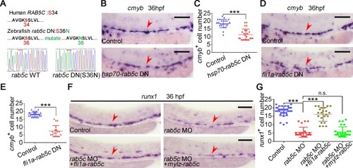
(A) Amino acid sequence is conserved for GTP-binding pockets of the Rab5c proteins between human and zebrafish. The amino acid of zebrafish in red was mutated to generate DN Rab protein by affecting GTP/GDP affinity. (B) Expression of cmyb in control and Rab5c inhibition groups examined by WISH. rab5c DN overexpression was carried out by hsp70-GFP-rab5c DN HS at 20 hpf. Scale bar, 100 μm. (C) Quantification of the cmyb positive cells. Error bars, mean ± SD, ***P < 0.001. (D) WISH analysis shows that cmyb is decreased in embryos of rab5c DN overexpression driven by a fli1a promoter. Scale bar, 100 μm. (E) Quantification of the cmyb positive cells. Error bars, mean ± SD, ***P < 0.001. (F) Endothelial, but not somitic, Rab5c overexpression has a HSPC rescue. Scale bar, 100 μm. (G) Quantification of the runx1 positive cells. Error bars, mean ± SD, ***P < 0.001. The P values in this figure were calculated by Student t test. The underlying data in this figure can be found in S1 Data. DN, dominant-negative; EC, endothelial cell; GDP, guanosine diphosphate; GTP, guanosine triphosphate; HE, hemogenic endothelium; hpf, hours post fertilization; HS, heat shock; HSPC, hematopoietic stem and progenitor cell; n.s., nonsignificant; WISH, whole-mount in situ hybridization; WT, wild type .

Rab5c-regulated endocytic trafficking is important for HE specification through Notch signaling.

(A) The fli1a+ ECs were sorted from the trunk region of the control and the rab5c morphants at 26 hpf for RNA-seq. The GO analysis showing the enrichment of down-regulated signaling pathways in rab5c morphants. (B) GO analysis showing the enrichment of up-regulated signaling pathways in rab5c morphants. (C) Protein level of NICD in the control and the rab5c morphants at 26 hpf examined by WB. (D) Quantification and statistical analysis of WB. Quantification of protein level using gray analysis (Gel-Pro analyzer). Error bars, mean ± SD, ***P < 0.001. (E) Confocal imaging shows the fli1a+tp1+ Notch-active ECs in the VDA region (box area) of the control and the rab5c morphants. Scale bar, 100 μm. (F) Quantification of fli1a+tp1+ Notch-active ECs. Error bars, mean ± SD, ***P < 0.001. (G) Expression of Notch downstream genes ephrinB2a and gata2b in the control and the rab5c morphants at 26 hpf examined by WISH. Scale bar, 100 μm. (H) Statistical analysis of the WISH. Error bars, mean ± SD, ***P < 0.001. (I) Quantification of the gata2b positive cells. Error bars, mean ± SD, ***P < 0.001. (J) Relative mRNA level of Notch signaling downstream genes hey1, hey2 in the control and the rab5c morphants at 26 hpf examined by qRT-PCR. Error bars, mean ± SD, ***P < 0.001. (K) Expression of ephrinB2a in control and Rab5c inhibition group examined by WISH. rab5c DN overexpression was carried out by hsp70-GFP-rab5c DN HS at 20 hpf. Scale bar, 100 μm. (L) Statistical analysis of the WISH. Error bars, mean ± SD, ***P < 0.001. (M) WISH shows that runx1 expression in rab5c morphants is partially rescued by NICD overexpression through fli1a-NICD. Scale bar, 100 μm. (N) Quantification of the runx1 positive cells. Error bars, mean ± SD, ***P < 0.001. (O) Control plasmid or pCS2-rab5c DN transfected 293T cells were immunostained with antibodies against endogenous JAG1 (green) and EEA1 (red). Scale bar, 10 μm. (P) Quantification of co-localization of JAG1 with EEA1 using Manders’ coefficient (ImageJ). n = 14 cells. Error bars, mean ± SD, ***P < 0.001. (Q) Control plasmid or pCS2-rab5c DN transfected 293T cells were immunostained with antibodies against endogenous DLL4 (green) and EEA1 (red). Scale bar, 10 μm. (R) Quantification of co-localization of DLL4 with EEA1 using Manders’ coefficient. n = 14 cells. Error bars, mean ± SD, ***P < 0.001. (S) Control plasmid or pCS2-rab5c DN transfected 293T cells were immunostained with antibodies against endogenous NOTCH1 (green) and EEA1 (red). Scale bar, 10 μm. (T) Quantification of co-localization of NOTCH1 with EEA1 using Manders’ coefficient. n = 14 cells. Error bars, mean ± SD, ***P < 0.001. The P values in this figure were calculated by Student t test. The underlying data in this figure can be found in S1 Data. DN, dominant-negative; EC, endothelial cell; GO, gene ontology; HE, hemogenic endothelium; hpf, hours post fertilization; HS, heat shock; NICD, Notch intracellular domain; qRT-PCR, quantitative reverse-transcription PCR; RNA-seq, RNA sequencing; VDA, ventral wall of the dorsal aorta; WB, western blot; WISH, whole-mount in situ hybridization.

Rab5c is required for HE survival through AKT signaling.

(A) Protein level of p-Akt and total Akt in the control and the rab5c morphants examined by WB. (B) Quantification of protein level using gray analysis (Gel-Pro analyzer). Error bars, mean ± SD, ***P < 0.001. (C) TUNEL assay shows that there are more apoptotic HE cells (yellow) in the VDA region (box area) of rab5c morphants compared with the control. Injection of tp53 MO just slightly reduces apoptotic HE cell number in rab5c morphants. Scale bar, 100 μm. (D) Quantification of TUNEL+ HE cells. Error bars, mean ± SD, *P < 0.05, ***P < 0.001. (E) HSPC rescue of rab5c morphants with CA form AKT2 mRNA. Scale bar, 100 μm. (F) Quantification of the runx1 positive cells. Error bars, mean ± SD, ***P < 0.001. (G) Co-overexpression of NICD and AKT2 CA shows a more efficient rescue effect than NICD overexpression alone. Scale bar, 100 μm. (H) Quantification of the runx1 positive cells. Error bars, mean ± SD, ***P < 0.001. (I) Confocal imaging shows partial co-localization of Appl1 and Rab5c in 293T cells co-transfected with pCS2-mCherry-appl1 and pCS2-GFP-rab5c constructs. Scale bar, 10 μm. (J) 293T cells were co-transfected with flag-tagged rab5c and myc-tagged appl1 constructs. Cell lysate was subjected to IP using anti-flag beads followed by WB analysis. (K) Quantification of protein level using gray analysis (Gel-Pro analyzer). The ratios of Appl1-myc co-IP relative to input were calculated. Error bars, mean ± SD, ***P < 0.001. (L) Control plasmid or pCS2-rab5c DN transfected 293T cells were immunostained with antibodies against endogenous APPL1 (green) and AKT (red). Scale bar, 10 μm. (M) Quantification of co-localization of APPL1 with AKT using Manders’ coefficient (ImageJ). n = 14 cells. Error bars, mean ± SD, ***P < 0.001. (N) Control plasmid or pCS2-rab5c DN transfected 293T cells were immunostained with antibodies against endogenous PIK3CA (green) and AKT (red). Scale bar, 10 μm. (O) Quantification of co-localization of PIK3CA with AKT using Manders’ coefficient. n = 14 cells. Error bars, mean ± SD, ***P < 0.001. The P values in this figure were calculated by Student t test. The underlying data in this figure can be found in S1 Data. CA, constitutively active; DN, dominant-negative; GFP, green fluorescent protein; HE, hemogenic endothelium; HSPC, hematopoietic stem and progenitor cell; IP, immunoprecipitation; MO, morpholino; NICD, Notch intracellular domain; n.s., nonsignificant; p-Akt, phosphorylated Akt; PIK3CA, catalytic subunit PI3K-alpha; TUNEL, terminal-deoxynucleoitidyl transferase mediated nick end labeling; VDA, ventral wall of the dorsal aorta; WB, western blot .

Rab5c overactivation leads to HSPC production defect.

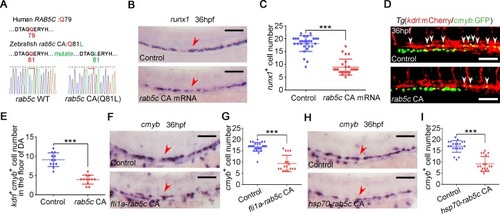
(A) Amino acid sequence is conserved for GTP hydrolysis of the Rab5c proteins between human and zebrafish. The amino acid of zebrafish in red was mutated to generate CA Rab protein by affecting GTPase activity. (B) WISH analysis shows that runx1 is decreased in rab5c CA mRNA overexpressed embryos. Scale bar, 100 μm. (C) Quantification of the runx1 positive cells. Error bars, mean ± SD, ***P < 0.001. (D) Confocal imaging shows the kdrl+cmyb+ definitive hematopoietic precursors in the VDA region of control and Rab5c CA overexpression group at 36 hpf. White arrowheads denote precursors. Scale bar, 100 μm. (E) Quantification of kdrl+cmyb+ cells. Error bars, mean ± SD, ***P < 0.001. (F) WISH analysis shows that cmyb is decreased in embryos of rab5c CA overexpression driven by fli1a promoter. Scale bar, 100 μm. (G) Quantification of the cmyb positive cells. Error bars, mean ± SD, ***P < 0.001. (H) Expression of cmyb in control and Rab5c CA overexpression group examined by WISH. hsp70-GFP-rab5c CA construct injected embryos were HS at 20 hpf for Rab5c CA overexpression. Scale bar, 100 μm. (I) Quantification of the cmyb positive cells. Error bars, mean ± SD, ***P < 0.001. The P values in this figure were calculated by Student t test. The underlying data in this figure can be found in S1 Data. CA, constitutively active; GFP, green fluorescent protein; GTP, guanosine triphosphate; hpf, hours post fertilization; HS, heat shock; HSPC, hematopoietic stem and progenitor cell; VDA, ventral wall of the dorsal aorta; WISH, whole-mount in situ hybridization .

Excessive endocytic trafficking mediated by Rab5c overactivation impairs Notch signaling during HSPC specification.

(A) Protein level of NICD in control and rab5c CA group at 26 hpf examined by WB. (B) Quantification of protein level using gray analysis. Error bars, mean ± SD, ***P < 0.001. (C) Confocal imaging shows the fli1a+tp1+ Notch-active ECs in the VDA region (box area) of control and rab5c CA group. Scale bar, 100 μm. (D) Quantification of fli1a+tp1+ Notch-active ECs. Error bars, mean ± SD, ***P < 0.001. (E) Expression of Notch downstream genes ephrinB2a and gata2b in control and rab5c CA group at 26 hpf examined by WISH. Scale bar, 100 μm. (F) Statistical analysis of the WISH. Error bars, mean ± SD, ***P < 0.001. (G) Quantification of the gata2b positive cells. Error bars, mean ± SD, ***P < 0.001. (H) Relative mRNA level of Notch signaling downstream genes hey1, hey2 in the control and the rab5c CA group at 26 hpf examined by qRT-PCR. Error bars, mean ± SD, ***P < 0.001. (I) WISH analysis shows that runx1 expression in Rab5c CA HS overexpression group is partially rescued by NICD overexpression through fli1a-NICD. Scale bar, 100 μm. (J) Quantification of the runx1 positive cells. Error bars, mean ± SD, ***P < 0.001. (K) Control plasmid or pCS2-rab5c CA transfected 293T cells were immunostained with antibodies against endogenous JAG1 (green) and EEA1 (red). Scale bar, 10 μm. (L) Quantification of co-localization of JAG1 with EEA1 using Manders’ coefficient (ImageJ). n = 13 cells. Error bars, mean ± SD, ***P < 0.001. (M) Control plasmid or pCS2-rab5c CA transfected 293T cells were immunostained with antibodies against endogenous DLL4 (green) and EEA1 (red). Scale bar, 10 μm. (N) Quantification of co-localization of DLL4 with EEA1 using Manders’ coefficient. n = 12 cells. Error bars, mean ± SD, ***P < 0.001. (O) Control plasmid or pCS2-rab5c CA transfected 293T cells were immunostained with antibodies against endogenous NOTCH1 (green) and EEA1 (red). Scale bar, 10 μm. (P) Quantification of co-localization of NOTCH1 with EEA1 using Manders’ coefficient. n = 14 cells. Error bars, mean ± SD, ***P < 0.001. The P values in this figure were calculated by Student t test. The underlying data in this figure can be found in S1 Data. CA, constitutively active; EC, endothelial cell; hpf, hours post fertilization; HS, heat shock; HSPC, hematopoietic stem and progenitor cell; NICD, Notch intracellular domain; qRT-PCR, quantitative reverse-transcription PCR; VDA, ventral wall of the dorsal aorta; WB, western blot; WISH, whole-mount in situ hybridization.

Acknowledgments
This image is the copyrighted work of the attributed author or publisher, and ZFIN has permission only to display this image to its users. Additional permissions should be obtained from the applicable author or publisher of the image. Full text @ PLoS Biol.