IMAGE

Fig 6

ID
ZDB-IMAGE-200501-46
Source
Figures for Heng et al., 2020
Image
Figure Caption

Fig 6 Rab5c overactivation leads to HSPC production defect.

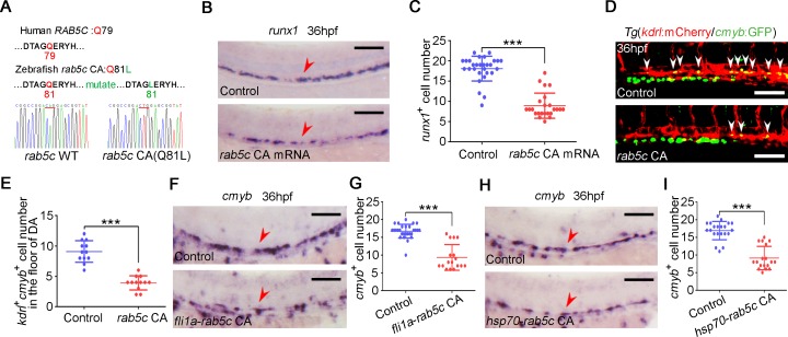
(A) Amino acid sequence is conserved for GTP hydrolysis of the Rab5c proteins between human and zebrafish. The amino acid of zebrafish in red was mutated to generate CA Rab protein by affecting GTPase activity. (B) WISH analysis shows that runx1 is decreased in rab5c CA mRNA overexpressed embryos. Scale bar, 100 μm. (C) Quantification of the runx1 positive cells. Error bars, mean ± SD, ***P < 0.001. (D) Confocal imaging shows the kdrl+cmyb+ definitive hematopoietic precursors in the VDA region of control and Rab5c CA overexpression group at 36 hpf. White arrowheads denote precursors. Scale bar, 100 μm. (E) Quantification of kdrl+cmyb+ cells. Error bars, mean ± SD, ***P < 0.001. (F) WISH analysis shows that cmyb is decreased in embryos of rab5c CA overexpression driven by fli1a promoter. Scale bar, 100 μm. (G) Quantification of the cmyb positive cells. Error bars, mean ± SD, ***P < 0.001. (H) Expression of cmyb in control and Rab5c CA overexpression group examined by WISH. hsp70-GFP-rab5c CA construct injected embryos were HS at 20 hpf for Rab5c CA overexpression. Scale bar, 100 μm. (I) Quantification of the cmyb positive cells. Error bars, mean ± SD, ***P < 0.001. The P values in this figure were calculated by Student t test. The underlying data in this figure can be found in S1 Data. CA, constitutively active; GFP, green fluorescent protein; GTP, guanosine triphosphate; hpf, hours post fertilization; HS, heat shock; HSPC, hematopoietic stem and progenitor cell; VDA, ventral wall of the dorsal aorta; WISH, whole-mount in situ hybridization .

Acknowledgments
This image is the copyrighted work of the attributed author or publisher, and ZFIN has permission only to display this image to its users. Additional permissions should be obtained from the applicable author or publisher of the image. Full text @ PLoS Biol.