
- Title
-
Transcriptomic profile of early zebrafish PGCs by single cell sequencing
- Authors
- Zhang, X., Li, X., Li, R., Zhang, Y., Li, Y., Li, S.
- Source
- Full text @ PLoS One
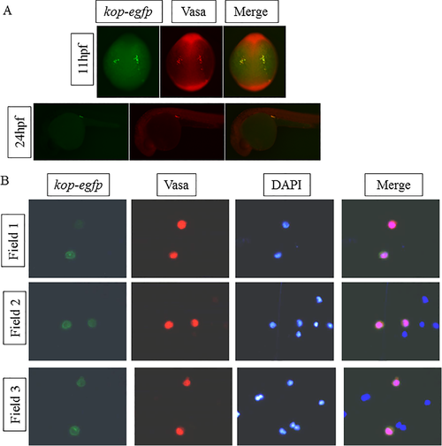
Confirmation of the zebrafish PGCs by immunostaining with Vasa antibody. (A) Whole mount immunostaining with Vasa antibody. Cells containing Vasa proteins can give out red fluorescence (second column). (B) Digested cells from zebrafish gonad at 24 hpf stained by Vasa antibody (second column) and DAPI (third column). Field 1, Field 2 and Field 3 mean three different fields of the glass slide under the microscope. EXPRESSION / LABELING:
|

ZFIN is incorporating published figure images and captions as part of an ongoing project. Figures from some publications have not yet been curated, or are not available for display because of copyright restrictions. |

Images for the zebrafish embryos in this study.White arrows pointing to cells emitting green fluorescence means PGCs. Images of the up panel are taken under bright light, and images of the down panel are taken under fluorescent light. |