
- Title
-
Fertility Enhancement but Premature Ovarian Failure in esr1-Deficient Female Zebrafish.
- Authors
- Chen, Y., Tang, H., Wang, L., He, J., Guo, Y., Liu, Y., Liu, X., Lin, H.
- Source
- Full text @ Front Endocrinol (Lausanne)

Morphological and histological analyses of esr1 mutant female zebrafish. (A) Body length of esr1+/+ and esr1−/− female zebrafish. (B) Body weight of esr1+/+ and esr1−/− female zebrafish. (C) GSI of esr1+/+ and esr1−/− female zebrafish. (D–M) Gross morphology of female zebrafish. (D′-G′) Ovaries of esr1+/+ and esr1 mutant were normal at 60 and 90 dpf. (H′-M′) Part of esr1 mutant ovaries of at 120 dpf and all esr1 mutant ovaries were degenerated by the 180 dpf. In these degenerated ovaries, MV and FG follicles were absent and PG follicles, PV follicles and atretic follicles (asterisk) were found. PG, primary growth follicle; PV, pre-vitelligenic follicle; EV, early-vitelligenic follicle; MV, middle-vetelligenic follicle; LV, late-vitelligenic follicle; FG, full grown follicle; *, atretic follicle. PHENOTYPE:
|

ZFIN is incorporating published figure images and captions as part of an ongoing project. Figures from some publications have not yet been curated, or are not available for display because of copyright restrictions. PHENOTYPE:
|
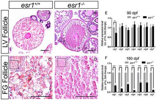
Vitellogenesis in esr1 deficient female zebrafish. (A) LV stage follicles were filled with vitellogenic granules in wild-type zebrafish. (B) Vitellogenic granules were found in the lateral part of LV stage follicles in esr1 mutants. (C) FG stage follicles were filled with big full vitellogenic granules in wild-types. (D) Only small vitellogenic granules were found in FG stage follicles of esr1 mutants. (E,F) Expression level of seven vtg genes at 90 dpf (E) and 180 dpf (F). The mRNA levels were normalized to ef1a. Average mRNA levels in ovaries of wild-types were defined as 1.0. (*P < 0.05; ***P < 0.001; Mean ± SEM). |
|
ZFIN is incorporating published figure images and captions as part of an ongoing project. Figures from some publications have not yet been curated, or are not available for display because of copyright restrictions. |
|
ZFIN is incorporating published figure images and captions as part of an ongoing project. Figures from some publications have not yet been curated, or are not available for display because of copyright restrictions. |
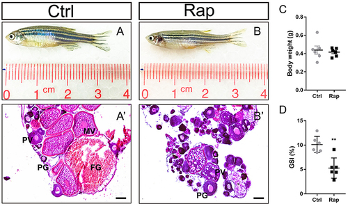
Morphology and ovarian histology of rapamycin treated zebrafish (A,B) Gross morphology of control and rapamycin treated zebrafish. (A′,B′) In rapamycin treated zebrafish, only PG follicles, PV follicles and atretic follicles (asterisk) were found in the ovaries. (C,D) Body weight (C,D) GSI of control and rapamycin treated zebrafish. (*P < 0.05; **P < 0.01; ***P < 0.001; Mean ± SEM). PG, primary growth follicle; PV, pre-vitelligenic follicle; *, atretic follicle. PHENOTYPE:
|