
Image
|
Figure Caption
Fig. 7
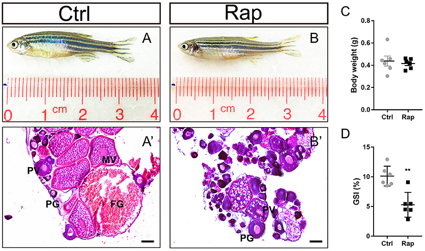
Morphology and ovarian histology of rapamycin treated zebrafish (A,B) Gross morphology of control and rapamycin treated zebrafish. (A′,B′) In rapamycin treated zebrafish, only PG follicles, PV follicles and atretic follicles (asterisk) were found in the ovaries. (C,D) Body weight (C,D) GSI of control and rapamycin treated zebrafish. (*P < 0.05; **P < 0.01; ***P < 0.001; Mean ± SEM). PG, primary growth follicle; PV, pre-vitelligenic follicle; *, atretic follicle.
Figure Data
Acknowledgments
This image is the copyrighted work of the attributed author or publisher, and
ZFIN has permission only to display this image to its users.
Additional permissions should be obtained from the applicable author or publisher of the image.
Full text @ Front Endocrinol (Lausanne)