- Title
-
Distinct Neuroblastoma-associated Alterations of PHOX2B Impair Sympathetic Neuronal Differentiation in Zebrafish Models
- Authors
- Pei, D., Luther, W., Wang, W., Paw, B.H., Stewart, R.A., and George, R.E.
- Source
- Full text @ PLoS Genet.
|
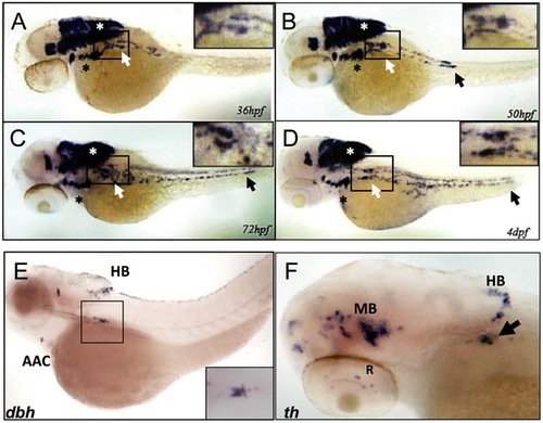
phox2b is expressed in the SCG, a marker of the peripheral sympathetic nervous system in the zebrafish. (A–D) Whole-mount in situ hybridization (ISH) for phox2b expression in wild-type embryos at the indicated time points. phox2b expression is seen in cells of the prospective superior cervical ganglion (SCG; white arrows, boxed) at 36 hpf (A), which start to extend caudally to form the sympathetic chain. These cells are identified as sympathetic neuronal cells by their expression of dbh and th (E, F). Expression is also seen in the brachial arches (black asterisk) and hindbrain (white asterisk). By 50 hpf, phox2b expression is seen in the enteric precursors (B–D, black arrow). Expression continues in two parallel rows caudally, encompassing the sympathetic chain until 4 dpf. (E) Lateral view (cranial to the left) of a 4-dpf zebrafish embryo analyzed by whole-mount ISH for dbh expression, which is seen in the SCG (black square), the arch associated complex (AAC) also derived from the neural crest, and the hindbrain (HB). Inset shows enlarged dorsal view of the SCG. (F) Whole-mount ISH of 4-dpf embryo analyzed for th expression. Aggregates of cells expressing th are seen in the SCG (arrow) as well as in the midbrain (MB), HB and retina (R). |
|
phox2b-deficient embryos show impaired differentiation of sympathetic neurons in the SCG. (A–F) Lateral/oblique views of 4-dpf embryos after whole-mount ISH for th (A–C) and dbh (D–F) in control and phox2b–deficient embryos. Arrows indicate the SCG. Knockdown of phox2b by injection of a splice MO (4 ng) (MOsplice) inhibits the expression of th and dbh (B, E) which is rescued by coexpression of human PHOX2B mRNA (10 ng/μl) (C, F). Relative intensity levels of th (G) and dbh (H) expression in embryos injected with phox2b MOs that inhibit translation (MOATG) or splicing (MOsplice). Mismatched control MO (MOmm) and PHOX2B mRNA-rescue (MOsplice/PHOX2B) are also shown. Data are presented as means ± SD (***P<0.001; **P<0.01; n = 15 for each group). |
|
The neuroblastoma-associated 676delG and K155X PHOX2B variants cause decreased terminal differentiation in the SCG. Whole-mount ISH for th (A–F) and dbh (G–L) expression in 3-dpf embryos in which human neuroblastoma-derived mutations were overexpressed (lateral views are shown). The area encompassing the SCG (boxed) is shown enlarged to the right of each panel. Capped mRNA (100 ng/μl) for wild-type (WT) human PHOX2B and the R100L, 676delG, K155X mutations were injected into one-cell embryos. CT, control water-injected. Relative intensity levels of th (M) and dbh (N) expression in the embryos depicted in panels A–F and G–L respectively. Data are presented as means ± SD (*P<0.01 vs. control-injected embryos; n = 6 per group). Whole-mount ISH for th (O) and dbh (P) in 3-dpf embryos expressing the phox2b MO and PHOX2B K155X mutant mRNA (P2BMO+K155X). Arrow indicates the region of the SCG. EXPRESSION / LABELING:
PHENOTYPE:
|
|
Impaired differentiation in the SCG due to phox2b deficiency is not rescued by retinoic acid. (A–F) Dorsal views of 3-dpf embryos injected with phox2b MO (D–F) or mismatched control MO (A–C) and treated with increasing concentrations of 13–cis retinoic acid (RA). (G) Relative intensity measurements of th expression in the SCG of embryos injected with various MOs and treated with different concentrations of RA. ATGK1, translation-blocking phox2b MO; P2BT2, splice blocking phox2b MO. (H–O) Whole-mount ISH of 3-dpf embryos in which the specified RNAs were overexpressed and analyzed for th expression following exposure to RA. Capped mRNA (100 ng/µl) for wild-type (WT) human PHOX2B, and the R100L, 676delG, K155X mutations were injected into embryos at the one-cell stage. (P) Quantification of the relative intensity of th staining in the embryos depicted in H–O. Data are presented as means ± SD (*P<0.05; n = 10 per group). EXPRESSION / LABELING:
|
|
Deficiency of Phox2b protein due to either MO knockdown or overexpression of PHOX2B variants leads to increased phox2b and ascl1 RNA expression in the SCG. (A–D) Dorsal views (cranial to the left) of 4-dpf embryos expressing a phox2b ATG MO (B,D) or mismatched control MO (A,C), showing expression of phox2b (A, B) and ascl1 (C, D) as determined by whole-mount ISH. Insets depict an enlarged view of the area of the SCG. (E–I) Area of the SCG is shown in 4-dpf embryos (lateral view, cranial to the left) in which capped mRNA (100 ng/μl) for wild-type (WT) human PHOX2B (F) and the indicated variants (G–I) was injected and ISH performed for ascl1. CT, control, water injected. (J, K) Whole-mount ISH in control vs. phox2b MO-injected embryos double labeled with phox2a (blue) and th (red) riboprobes. Arrows point to the SCG. EXPRESSION / LABELING:
|
|
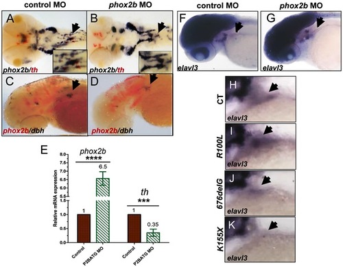
Phox2b deficiency causes arrest of SCG cells at an undifferentiated stage. (A,B) Dorsal views of ISH with FITC-labeled th (red) and digoxigenin-labeled phox2b (blue) riboprobes on 4-dpf embryos in which phox2b expression was abrogated by MO knockdown. Arrows point to the SCG. Insets show enlarged views of the SCG. (C, D) Lateral views of ISH with FITC-labeled phox2b (red) and digoxigenin-labeled dbh (blue) riboprobes in MO-injected (D) and control (C) embryos. (E) Quantitative real time-PCR analysis comparing phox2b mRNA expression levels in control vs. phox2b MO-injected (P2BATG MO) embryos. Data are presented as means ± SD (****P<0.0001, ***P<0.001; n = 15 per group). (F,G) Whole-mount ISH for elavl3 in the area of the SCG (arrow) in phox2b MO-injected (G) compared to control MO-injected embryos at 4-dpf (F). (H–K) Lateral views of the SCG in 4-dpf embryos overexpressing the indicated RNAs analyzed for elavl3 expression by whole-mount ISH. |
|
phox2b, but not ascl1, is indispensable for sympathetic neuronal differentiation. (A–F) Whole-mount ISH of 3-dpf embryos for th and dbh following ascl1 MO knockdown. Lateral views depict a minimal decrease in expression of th (A,B) and dbh (D,E) in ascl1 MO-injected embryos compared to control MO-injected (A,D) embryos. Simultaneous knockdown of both ascl1 and phox2b led to a significant decrease in expression of th and dbh in the SCG (C,F). (G) Relative intensity levels of dbh-staining in the SCG of embryos expressing ascl1 MO, phox2b MO or the combination of the two. Data are presented as means ± SEM (***P<0.001; n = 15 per group). (H–M) Whole-mount ISH for expression of ascl (H–J) and phox2b (K–M) in embryos in which ascl1 expression was abrogated by MO knockdown, either singly (I, L) or in combination with phox2b (J, M). (N,O) Whole-mount ISH for phox2a expression in 4-dpf embryos in which ascl1 expression was abrogated by MO knockdown. Arrows point to region of SCG. EXPRESSION / LABELING:
PHENOTYPE:
|


