- Title
-
Analysis of Sphingosine-1-phosphate signaling mutants reveals endodermal requirements for the growth but not dorsoventral patterning of jaw skeletal precursors
- Authors
- Balczerski, B., Matsutani, M., Castillo, P., Osborne, N., Stainier, D.Y., and Crump, J.G.
- Source
- Full text @ Dev. Biol.
|
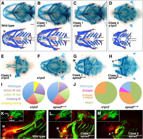
Jaw skeletal defects in s1pr2 and spns2b1110 mutants. (A–H) Ventral views show head skeletons of 6 dpf zebrafish larvae stained for cartilage (blue) and bone (red) in (A–F) and cartilage only in (G, H). Bottom schematics in (A–D) show facial cartilage (dark blue), neurocranial cartilage (light blue), and bone and teeth (red). Meckel′s (M), pterygoid process (Ptp), and trabecular (Tr) cartilages are indicated. s1pr2 and spns2b1110 larvae variably displayed ectopic (e) midline cartilage (Class 1: B, C, G) and/or graded reductions of jaw and anterior neurocranial skeleton (Class 2: D, E, H). In addition, some genotypically mutant s1pr2 and spns2b1110 larvae displayed a normally patterned jaw (F). (I and J) Pie charts show proportions of s1pr2 (n = 32) and spns2b1110 (n = 30) larvae showing progressive reductions of jaw skeleton (I, scored for each side) and ectopic cartilage and/or heart defects (J, scored for each animal). Minor M reduction refers to losses of less than 50% of the cartilage (e.g. C, G) and major M reduction losses greater than 50% (e.g. top of D). More severe categories include missing M (e.g. bottom D) and missing M + Pq (e.g. E). Wild-type siblings never displayed defects. (K–M) sox10:kikGR imaging of the mandibular and hyoid arches at 30 hpf (insets) and the resulting skeletal derivatives at 5 dpf. Photoconversion of mandibular (mand) prominence CNCCs (red) resulted in labeling of Meckel′s (M) cartilage and surrounding mesenchyme in wild types (n = 3/3). In spns2b1110; sox10:kikGR larvae, mandibular CNCCs generated either malformed M and ectopic (e) midline cartilages (L, n = 3/9) or very reduced remnants (“X”) (M, n = 6/9). Max, maxillary prominence. Scale bars = 50 μm. |
|
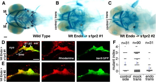
Wild-type endoderm rescues lower jaw development in s1pr2 mutants. (A–C) Ventral views of 6 dpf head skeletons. Unilateral transplantation of wild-type endoderm precursors into s1pr2 mutants rescued the loss (B) or reduction (C) of lower jaw M cartilage seen in control sides not receiving transplants. (D and E) The same two examples imaged at 30 hpf to show the contribution of wild-type her5:GFP+ (green) rhodamine-dextran+ (red) donor endoderm cells to the anterior medial endoderm (ame) and the first and second pouches (p1 and p2) of the host arches. (F) For quantification of lower jaw rescue, we devised a mutant index: 0, wild type; 1, minor M reduction; 2, major M reduction; 3, M missing; 4, M and Pq missing. Compared to s1pr2 mutants receiving mock transplants, mutant sides receiving endoderm transplants (but not the contralateral control sides of the same animals) had partial restoration of lower jaw cartilage. Individual data points are plotted, and bars show standard error of the mean. Asterisks denote the two groups with statistical differences in average jaw skeletal defects. Scale bars = 50 μm. |
|
Facial ectoderm gene expression in endoderm mutants. (A–T) In situ hybridizations at 36 hpf. In wild types, pitx2ca (A), shha (E), and fgf8a (I) are expressed in oral ectoderm and edn1 (M) and bmp4 (Q) in aboral ectoderm. pitx2ca expression in the oral ectoderm (arrowheads) is present but shifted anteriorly in sox32 (B, n = 6/6), s1pr2 (C, n = 11/11), and spns2b1110 (D, n = 4/4) embryos. The expression of shha in the oral ectoderm is lost in all mutants, whereas endoderm expression (arrows) is absent in sox32 mutants (F, n = 7/7), variably posteriorly displaced (G, n = 10/15) or disorganized (see 4, n = 3/15) in s1pr2 mutants, and variably posteriorly displaced (H, n = 6/7) or disorganized (not shown, n = 1/7) in spns2b1110 mutants. fgf8a expression is reduced in sox32 mutants (J, n = 6/6), reduced (K, n = 4/10) or disorganized (not shown, n = 4/10) in s1pr2 mutants, and reduced (not shown, n = 13/27) or disorganized (L, n = 14/27) in spns2b1110 mutants. edn1 expression in the first two arches (white lines) is present but disorganized in sox32 (N, n = 8/8), s1pr2 (O, n = 6/6), and spns2b1110 (P, n = 5/5) mutants. bmp4 expression is also disorganized in sox32 (R, n = 6/6), s1pr2 (S, n = 7/7), and spns2b1110 (T, n = 5/5) mutants. Outlines of the oral ectoderm and first two pouches are shown for wild types. Scale bar = 50 μm. |
|
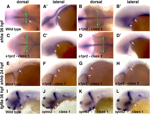
Variable disorganization of Shh-expressing endoderm and fgf8a ectodermal expression in s1pr2 and spns2b1110 mutants. (A–D) In situ hybridizations at 36 hpf show two classes of shha expression in s1pr2 mutants. When viewed dorsally Class 1 mutants have a wider shha endodermal expression domain (green arrows) than Class 2 mutants, and in lateral views of the same embryos Class 1 but not Class 2 mutants display ectopic shha expression (arrowheads) anterior to the normal expression domains (arrows). (E–H) In situ hybridizations at 24 hpf also show two classes of shhb expression in s1pr2 mutants, with Class I mutants (n = 3/8) showing ectopic anterior expression and Class 2 mutants (n = 5/8) displaying posterior truncation of the normal endodermal expression domain (arrows). (I–L) Several examples of spns2b1110 Class I mutants show fragmented expression of fgf8a expression in the oral ectoderm (arrowheads). Scale bar = 50 μm. |
|
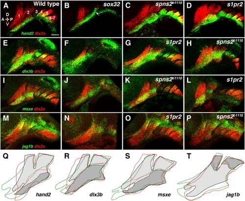
DV gene expression in endoderm mutants. (A–P) Double fluorescent in situ hybridizations show the expression of dlx2a (red) with hand2, dlx3b, msxe, or jag1b (green) in the pharyngeal arches (numbered) at 36 hpf. In wild types, hand2 (A) was restricted to the ventral-most domain, dlx3b (E) and msxe (I) to a DV-intermediate domain, and jag1b (M) to the dorsal arches. hand2 expression remained restricted to ventral arch CNCCs in sox32 embryos, although ectopic hand2 expression was seen in ectoderm overlying the second pouch (arrow) (B, n = 12/12). The hand2 expression domain remained ventrally restricted but was expanded in size in s1pr2 (D, n = 10/10), and spns2b1110 (C, n = 10/10) embryos. The DV-intermediate expression of dlx3b was normal in sox32 (F, n = 4/4), s1pr2 (G, n = 10/10), and spns2b1110 (H, n = 5/5) embryos, as was expression of msxe in sox32 (J, n = 7/7), s1pr2 (L, n = 11/11), and spns2b1110 (K, n = 5/5) embryos. jag1b expression was reduced but remained dorsal-restricted in sox32 embryos (N, n = 5/5) and was unaffected in s1pr2 (O, n = 5/5) and spns2b1110 (P, n = 6/6) embryos. (Q–T) Tracings of the dlx2a-expressing arches (solid lines) and the DV expression borders (dashed lines) of hand2 (Q), dlx3b (R), msxe (S), or jag1b (T) show slight expansion of hand2 expression but no change in dlx3b, msxe, or jag1b expression in two classes of s1pr2 and spns2b1110 mutants. For wild types, dlx2a expression is light gray, with DV gene expression domains in darker gray. The mandibular arch was expanded (green lines) in some embryos (C, G, K, O; 47% of s1pr2 and 53% of spns2b1110) and was abnormally shaped and/or anteriorly truncated (red lines) in other embryos (D, H, L, P; 47% of s1pr2 and 16% of spns2b1110). Scale bar = 50 μm. |
|
Shha misexpression partially rescues the jaw defects of sox32 mutants. (A–D) Ventral views of 6 dpf head skeletons. Compared to hsp70I:Gal4 controls subjected to the same heat-shock treatment (A), hs-Shha larvae developed ectopic Tr-like cartilages (arrowheads) (B, n = 10/10). Whereas facial skeleton never formed in non-hs-Shha sox32 siblings (C, n = 0/10), jaw and hyoid cartilage was partially restored and ectopic cartilage was still present in sox32; hs-Shha larvae (D, n = 4/4). Flat-mount preparations (insets in A and D) show that Meckel′s (M), palatoquadrate (Pq), and hyosymplectic (Hs) cartilages, but not the hyoid-arch-derived ceratohyal (Ch) cartilage, were dysmorphic yet present in sox32; hs-Shha larvae. (E–H) In situ hybridizations show fgf8a oral ectoderm expression (arrows) at 36 hpf. Compared to hsp70I:Gal4 controls (E), fgf8 expression was shifted dorsal-anteriorly in hs-Shha embryos (F, n = 8/9) and partially (n = 3/34) or severely reduced (n = 31/34) in sox32 mutants (G). Shha misexpression resulted in increased and dorsal-laterally shifted fgf8a expression in sox32; hs-Shha embryos (H, n = 11/12). (I–L) Confocal projections show the pharyngeal arches (numbered) of control fli1a:GFP (I, n = 3), hs-Shha; fli1a:GFP (J, n = 4), sox32; fli1a:GFP (K, n = 10), and sox32; hs-Shha; fli1a:GFP (L, n = 7) embryos at 30 hpf. In sox32 and sox32; hs-Shha embryos, the lack of endoderm resulted in arches 3–7 remaining as a single mass. White outlines show the maxillary (max), mandibular (mand), and hyoid domains for wild type. (M–T) Anti-pH3 staining marks proliferating cells (red, M–P), Lysotracker marks regions of cell death (red, Q–T), and fli1a:GFP labels CNCCs (green) of the mandibular (1) and hyoid (2) arches in control (M and Q), hs-Shha (N and R), sox32 (O and S), and sox32; hs-Shha (P and T) embryos at 36 hpf. n = 10 for each genotype. Scale bars = 50 μm. (U) Quantification of arch domain area and numbers of pH3- and Lysotracker-positive cells per arch area in control hsp70I:Gal4 (con), hs-Shha, sox32, and sox32; hs-Shha embryos. Brackets indicate comparisons showing statistical significance with a Tukey–Kramer HSD test (α = 0.05). PHENOTYPE:
|
Reprinted from Developmental Biology, 361(2), Balczerski, B., Matsutani, M., Castillo, P., Osborne, N., Stainier, D.Y., and Crump, J.G., Analysis of Sphingosine-1-phosphate signaling mutants reveals endodermal requirements for the growth but not dorsoventral patterning of jaw skeletal precursors, 230-241, Copyright (2012) with permission from Elsevier. Full text @ Dev. Biol.


