- Title
-
Genetic ablation of neural crest cell diversification
- Authors
- Arduini, B.L., Bosse, K.M., and Henion, P.D.
- Source
- Full text @ Development
|
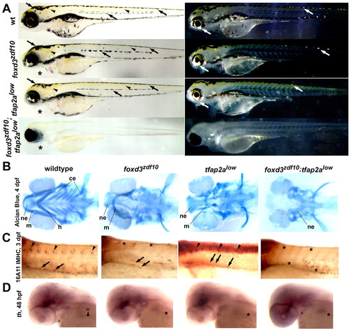
foxd3zdf10;tfap2alow double mutant embryos lack NC derivatives. (A) Live larvae at 4 days post-fertilization (dpf), lateral views with either transmitted (left) or reflected (right) light. Wild-type zebrafish exhibit three NC-derived pigment cell types: black melanophores (left, arrows), yellow xanthophores (left, arrowheads) and iridescent iridophores (right, white arrows). foxd3zdf10 and tfap2alow single mutants develop essentially normal pigment patterns by 4 dpf. Jaw structures protrude ventrally in both foxd3zdf10 and tfap2alow single mutants (asterisks). foxd3zdf10;tfap2alow double mutants completely lack NC-derived pigment cells except for occasional head xanthophores. Jaw structures appear to be missing (asterisk). (B) Craniofacial cartilage development revealed by Alcian Blue staining at 4 dpf, ventral views with anterior to the left. The wild-type larval head skeleton of zebrafish consists of the dorsal neurocranium (ne), as well as upper (mandibular, m) and lower (hyoid, h, and ceratobranchial, ce) jaw structures. In both foxd3zdf10 and tfap2alow single mutants, mandibular and hyoid structures are disorganized and the ceratobranchial elements are absent, whereas the neurocranium remains intact. Double mutants lack upper and lower jaw structures, and all but the most posterior portion of the neurocranium. (C) Immunostaining with 16A11 monoclonal (anti-Hu) antibody at 3 dpf, lateral views, anterior to the left. DRG neurons are found in each trunk segment of wild-type embryos (arrowheads) and enteric neurons populate the gut tube (arrows). DRG neurons are absent in foxd3zdf10 single mutants (asterisks) and present in reduced numbers in tfap2alow single mutants (arrowheads), whereas enteric neurons are reduced in number in both backgrounds (arrows). DRG and enteric neurons are absent in double mutant embryos (asterisks). (D) tyrosine hydroxylase (th) expression in sympathetic neurons at 48 hpf in wild-type embryos (arrowhead). Sympathetic neurons are absent in foxd3zdf10 and tfap2alow single mutants, as well as in double mutants (asterisks). PHENOTYPE:
|
|
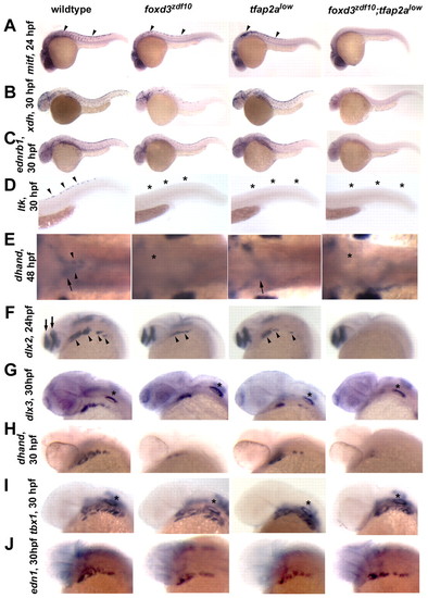
Failure of NC sublineage specification in foxd3zdf10;tfap2alow double mutants. (A-D) Lateral views with anterior to the left. (A) mitf expression at 24 hpf (arrowheads). foxd3zdf10;tfap2alow double mutant embryos completely lack NC-derived melanoblast mitf expression. (B) xanthine dehydrogenase (xdh), diagnostic of xanthophore precursors, at 30 hpf. (C) endothelin receptor B (ednrb1), presumed to be expressed by all chromatophore precursors, at 30 hpf and (D) leukocyte tyrosine kinase (ltk), reported to be expressed by iridophore precursors, at 30 hpf. (E) Dorsal views of dhand expression, anterior to left, ventral of the caudal hindbrain, at 48 hpf. Sympathetic neuron precursors are labeled in wild-type embryos (arrowheads), as are the more ventral and laterally localized enteric neuron precursors (arrows). In the majority of foxd3zdf10 embryos, both sympathetic and enteric neuron progenitors are absent (asterisk) (Stewart et al., 2006). In tfap2alow mutants, sympathetic neuron precursors are absent whereas a small number of enteric precursors are present (arrow) (Knight et al., 2003). In foxd3zdf10;tfap2alow double mutants, both sympathetic and enteric neuron precursors are absent (asterisk). (F-J) Lateral (F-I) and dorsolateral views (J), anterior to the left. (F) dlx2 expression at 24 hpf. dlx2 expression (arrowheads) is absent in the branchial arches of double mutant embryos, but is retained in the forebrain (arrows). (G) dlx3 expression at 30 hpf; (H) dhand expression at 30 hpf. NC expression of both genes is absent in double mutant embryos with dlx3 expression maintained in non-NC otic vesicle (asterisks). (I) tbx1 expression in the branchial arches (asterisk) at 30 hpf is diagnostic of the endodermal component of the arches. tbx1 expression is normal in all experimental embryos. (J) endothelin 1 (edn1) expression in the mesodermal component of the branchial arches at 30 hpf. Branchial mesoderm appears to develop normally in foxd3zdf10 and tfap2alow single mutants, as well as in foxd3zdf10;tfap2alow double mutant embryos. |
|
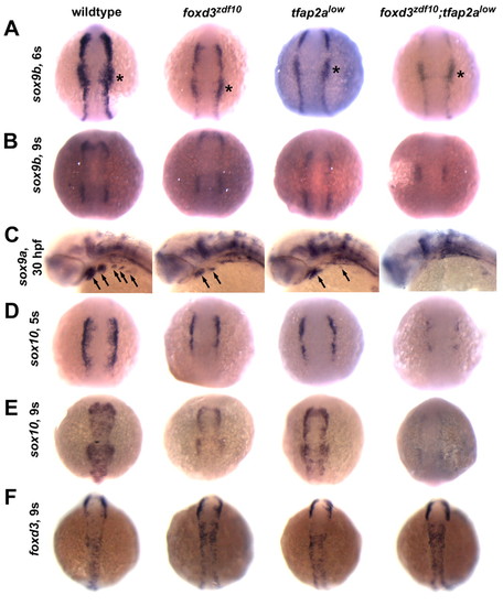
Defective expression of NC SoxE family genes in foxd3zdf10;tfap2alow double mutant embryos. (A,B,D-F) Dorsal views with anterior to the top of 6, 5 and 9 somite stage (s) embryos. (C) Lateral views with anterior to the left of 30 hpf embryos. (A) NC sox9b expression is severely depleted in foxd3zdf10;tfap2alow double mutants at the 6 somite stage, but is retained in the non-NC-derived otic placode (asterisks). (B) NC sox9b expression remains reduced in both single mutants, whereas NC expression is undetectable in double mutants by the 9 somite stage. (C) NC sox9a expression in the branchial arches (arrows) is absent in double mutant embryos. (D) There are slight reductions in NC sox10 expression in foxd3zdf10 and tfap2alow single mutants at the 5 somite stage, whereas in double mutant embryos NC sox10 expression is much more reduced. (E) By the 9 somite stage, NC sox10 expression is undetectable in double mutants. (F) In contrast to SoxE gene expression, NC foxd3 expression is maintained in foxd3zdf10;tfap2alow double mutant embryos, demonstrating that NC induction occurs. EXPRESSION / LABELING:
|
|
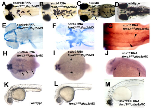
Misexpression of SoxE genes differentially rescues NC sublineage specification and differentiation in foxd3zdf10;tfap2aMO embryos. (A,E,H) sox9a and sox9b (sox9a/b) RNA co-injection with tfap2a morpholino (tfap2aMO) into foxd3zdf10 mutant embryos (foxd3zdf10-tfap2aMO). (A) Live 4 dpf larva, dorsal view, anterior to the left. Melanophore and xanthophore development is rescued in foxd3zdf10-tfap2aMO embryos co-injected with sox9a/b RNA (arrows; compare with wild-type embryo, D). (E) Alcian Blue staining of a 4 dpf larva, vental views with anterior to the left. Misexpression of sox9a/b in foxd3zdf10-tfap2aMO embryos results in rescue of the neurocranium, as well as of the mandibular and hyoid jaw structures. (H) dlx2 expression at 24 hpf, dorsal views, anterior to the left. Specification of NC craniofacial progenitors occurs in foxd3zdf10-tfap2aMO embryos co-injected with sox9a/b RNA, based on dlx2 expression (arrows). (B,F,G,I,J) foxd3zdf10-tfap2aMO embryos co-injected with sox10 RNA. (B) Live 4 dpf larva, dorsal view, anterior to the left. Consistent with a role in development of non-ectomesenchymal NC derivatives, sox10 misexpression rescues melanophore and xanthophore development in foxd3zdf10-tfap2aMO embryos (arrows; compare with wild-type embryo, D). (F) Alcian Blue staining of 4 dpf larva, ventral view, anterior to the left. sox10 misexpression does not rescue cranial cartilage structures in foxd3zdf10-tfap2aMO embryos. (G) 16A11 immunoreactivity, 3 dpf, dorsal view. Dorsal root ganglion neuron development is rescued by sox10 misexpression (arrowheads). (I) dlx2 expression at 24 hpf, dorsal view with anterior to the left. sox10 RNA injection also does not rescue NC dlx2 expression in foxd3zdf10-tfap2aMO embryos (asterisks), indicating that sox10 function cannot rescue the specification of NC precursors for craniofacial cartilages. (J) th expression at 48 hpf, lateral view, anterior to the left. sox10 misexpression rescues sympathetic neuron development (arrow). (C) Live 4 dpf larva, dorsal view, anterior to the left. Morpholino-mediated knockdown of p53 in foxd3zdf10-tfap2aMO embryos rescues melanophore, xanthophore (arrows; compare with wild-type embryo, D) and iridophore (not visible) development. (K-M) sox10-responsive NCCs persist in foxd3zdf10-tfap2aMO embryos. 33 hpf embryos, lateral view, anterior to left. (K) Wild-type embryo with abundant melanophores. (L) In foxd3zdf10-tfap2aMO embryos injected with a heat shock-inducible sox10 construct but not exposed to heat shock, melanophores fail to develop. (M) In foxd3zdf10-tfap2aMO embryos injected with a heat shock-inducible sox10 construct and heat shocked at 24 hpf, robust melanogenesis occurs. |
|
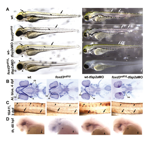
tfap2a morpholino-injected foxd3zdf10 mutant embryos precisely phenocopy foxd3zdf10;tfap2alow double mutants. Embryos obtained from foxd3zdf10 heterozygous matings were injected with tfap2a morpholino (tfap2aMO). Wild-type-tfap2aMO and foxd3zdf10-tfap2aMO phenocopy tfap2alow and foxd3zdf10;tfap2alow mutants, respectively, with 90% efficiency (n>1000). (A) Live larvae at 4 days post-fertilization (dpf), lateral views with either transmitted (left) or reflected (right) light. Compared with uninjected wild type, uninjected foxd3zdf10 mutant and wild-type tfap2aMO-injected siblings, foxd3zdf10-tfap2aMO larvae lack melanophores (left, arrows), xanthophores (left, arrowheads) and iridophores (right, white arrows), with the exception of occasional head xanthophores. (B) Alcian Blue staining at 4 dpf, ventral views, anterior to the left. All upper and lower jaw elements, as well as the anterior neurocranium, are absent in foxd3zdf10-tfap2aMO embryos. (C) 16A11 immunoreactivity, 3 dpf, lateral views with anterior to the left. DRG (arrowheads) and enteric neurons (arrows) are absent in mutant morphants. (D) th expression at 48 hpf, lateral views with anterior to the left. Sympathetic neurons (arrowheads) are absent in foxd3zdf10 mutants, tfap2a morphants and mutant morphants. |
|
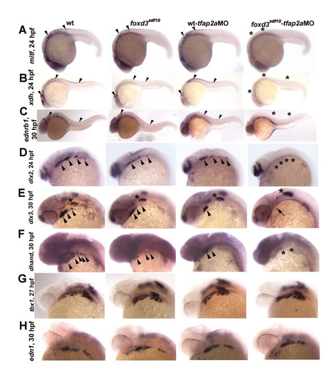
Neural crest sublineage specification fails to occur in foxd3zdf10-tfap2aMO embryos. (A-H) Lateral views with anterior to the left. (A) mitf expression at 24 hpf (arrowheads). Just as with foxd3zdf10;tfap2alow double mutant embryos, foxd3zdf10-tfap2aMO embryos completely lack neural crest (NC) mitf expression (asterisks). (B) xanthine dehydrogenase (xdh), diagnostic of xanthophore precursors, at 24 hpf (arrowheads); (C) endothelin receptor B (ednrb1), presumably expressed by all chromatophore precursors, at 30 hpf (arrowheads). NC expression of both genes is absent in mutant morphants (asterisks). (D) dlx2 expression at 24 hpf. dlx2 expression (arrowheads) is absent in the branchial arches of foxd3zdf10-tfap2aMO embryos (asterisks), but is retained in the forebrain. (E) dlx3 expression at 30 hpf (arrowheads). (F) dhand expression at 30 hpf (arrowheads). NC expression of both genes is absent in mutant morphants (arrow; double asterisks) with dlx3 expression maintained in non-NC-derived otic vesicle (asterisks). (G) tbx1 expression in the branchial arches at 27 hpf is diagnostic of the endodermal component of the arches. tbx1 expression is normal in all experimental embryos. (H) endothelin 1 (edn1) expression in the mesodermal component of the branchial arches at 30 hpf. Branchial mesoderm appears to be develop normally in foxd3zdf10 single mutant, tfap2aMO morphant and foxd3zdf10-tfap2aMO mutant-morphant embryos. |
|
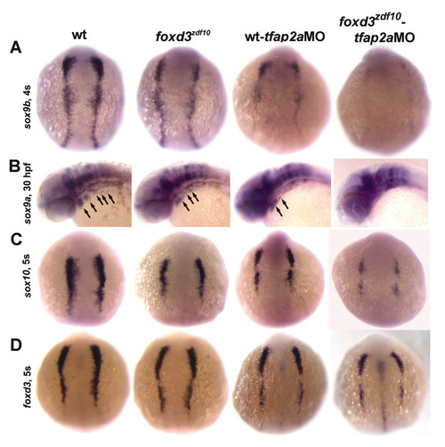
As in foxd3zdf10;tfap2alow double mutants, induction of NC SoxE gene expression is severely reduced in foxd3zdf10-tfap2aMO embryos. (A,C,D) Dorsal views, anterior up. (B) Lateral view, anterior to the left of a 30 hpf embryo. (A) At 4s, NC sox9b expression is virtually absent in mutant morphants. (C,D) At 5s, NC sox10 expression (C) is severely reduced in mutant morphants, whereas NC foxd3 expression (D) is retained. (B) NC sox9a expression in the pharyngeal arches (arrows) is undetectable in mutant morphants. |
|
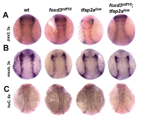
The neural plate border develops independent of foxd3 and tfap2a function. (A) pax3 expression at the 3 somite stage, dorsal views with anterior to the top. paired homeobox 3 (pax3) is expressed at the edge of the developing neural plate, and is required for induction of NC-specific gene expression. pax3 is robustly expressed in foxd3zdf10;tfap2alow double mutants, as well as in foxd3zdf10 and tfap2alow single mutant embryos. (B) msxb expression at the 3 somite stage, dorsal views with anterior to the top. msxb expression is required for proper size and positioning of the neural plate border. msxb expression is normal in foxd3zdf10;tfap2alow double mutant, foxd3zdf10 single mutant and tfap2alow single mutant embryos compared with wild-type siblings. (C) huC expression in 6 somite stage embryos. At this stage, primary motor neurons (more medial cells) and Rohon-Beard sensory neurons (bilateral stripes of cells just lateral to primary motorneurons) express huC. Both neuronal populations develop normally in both single mutants, as well as in double mutants. |

