- Title
-
Distinct functions of Wnt/{beta}-catenin signaling in KV development and cardiac asymmetry
- Authors
- Lin, X., and Xu, X.
- Source
- Full text @ Development
|
Wnt/β-catenin signaling regulates LR asymmetry. (A-E) APC is required for left-right patterning of zebrafish heart but not visceral organs. Wild-type sibling (A,C) and apc mutant (B,D) embryos were stained at 28 hpf (A,B) for cmlc2 and at 52 hpf (C,D) for trypsin. (E) Quantification of cardiac jogging and looping, and pancreas positioning. Shown are dorsal views with anterior to the top. (F-L) Activation and repression of Wnt/β-catenin signaling differentially affect cardiac and visceral organ laterality. (F-H) Representative images of cardiac looping as revealed by cmlc2 staining of 52 hpf embryos. Ventral views with anterior to the top. (I-K) Representative images of the positioning of visceral organs as revealed by foxa3 staining of 52 hpf embryos. Dorsal views with anterior to the top. l, liver; g, gut; p, pancreas. (L) Quantification of cardiac and visceral organ laterality. H, heart; G, gut and other visceral organs; n, total numbers of embryos scored. |
|
Wnt/β-catenin signaling is essential for the LR patterning function of KV. (A-F) charon expression was suppressed in wnt3 (D), wnt8 (E), and wnt3/wnt8 double (F) morphants, but not in embryos overexpressing wnt3 (B) or wnt8 (C) RNA, as compared with control embryos (A). Shown are dorsal views of tail bud regions at the 12-somite stage. (G-N) Cilia development in KV was regulated by Wnt signaling, as shown by anti-acetylated tubulin antibody staining of 10-somite staged embryos. Compared with controls (G; n=15), shorter KV cilia were observed in wnt3 morphants (J; n=15), wnt8 morphants (K; n=12), and wnt3/wnt8 double morphants (L; n=15), but not in embryos injected with wnt3 RNA (H; n=12) or wnt8 RNA (I; n=12), as summarized in M. Cilia numbers were increased in embryos overexpressing wnt3 (H) or wnt8 (I) RNA but remained the same in wnt3 (J), wnt8 (K) and wnt3/wnt8 double (L) morphants, as summarized in N. Data shown are means±s.d. *P<0.001, Student's t-test. EXPRESSION / LABELING:
PHENOTYPE:
|
|
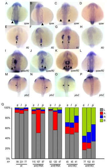
Activation of Wnt signaling ablates lefty2 expression in the heart without affecting the left-sided expression of spaw and pitx2. (A-P) Representative images of spaw expression in LPM (arrowheads in A-C,I-L), lefty2 expression in the cardiac field (arrows in E-G,I-K), and pitx2 expression in the posterior LPM (asterisks in M-O). Shown are dorsal views, with anterior to the top of 22- to 24-somite staged embryos. A tropomyosin probe was included in in situ hybridization to ensure embryos were at the proper stage, with the exception of pitx2 staining. (Q) Quantification of the percentages of asymmetric expression of spaw, lefty2 and pitx2. s, spaw; l, lefty2; p, pitx2. L, left side; A, absence; R, right side; B, bilateral; n, total numbers of embryos scored. PHENOTYPE:
|
|
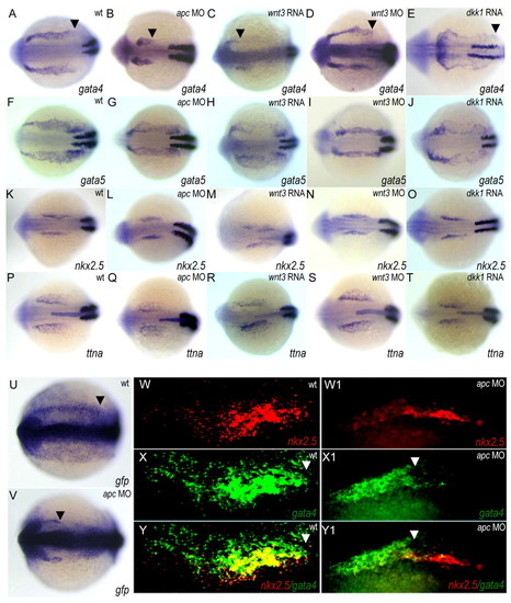
Extent of gata4 expression responds to Wnt/β-catenin signaling. (A-T) Wnt/β-catenin signaling regulates gata4 expression. (A-E) gata4 expression was restricted to the rostral ALPM in apc morphants (B) and in embryos injected with wnt3 RNA (C), was unaltered in wnt3 morphants (D), and was expanded caudally in dkk1-overexpressing embryos (E), compared with controls (A). (F-J) gata5 expression, (K-O) nkx2.5 expression, and (P-T) ttna expression were not affected. The embryos were co-stained with a myod probe to ensure the proper developmental stage. (U,V)Wnt signaling modulates gata4 expression at the transcriptional level. The expression of gfp in gata4::gfp transgenic fish was revealed by in situ hybridization using a gfp riboprobe. (W-Y1) Two-color fluorescent in situ hybridization showed that gata4 expression was partially depleted from the nkx2.5 expression domain and its lateral region in apc morphants (W1-Y1), compared with in controls (W-Y). Shown are dorsal views of 12-somite staged embryos with anterior to the left; only the right LPM is shown in W-Y1. Arrowheads indicate the posterior end of gata4 expression. EXPRESSION / LABELING:
|
|
Gata4 is required for cardiac LR patterning. (A) The gata4 (e2) morpholino efficiently disrupted mRNA splicing. RT-PCR on mRNA from morpholino-injected embryos amplified a 188-bp product (exon 2 skipping) and a 268-bp product (intron 2 retention); RT-PCR from control embryos only amplified a 454 bp product (normal splicing). Normally spliced gata4 mRNA in these morphants was 2.8% of the wild-type level as revealed by real-time PCR analysis. (B,C) Reducing Gata4 resulted in laterality defects, which can be rescued by co-injection with gata4 RNA in a dose-dependent manner. (B) Quantification of cardiac looping. (C) Quantification of asymmetric expression of spaw, lefty2 and pitx2. EXPRESSION / LABELING:
PHENOTYPE:
|
|
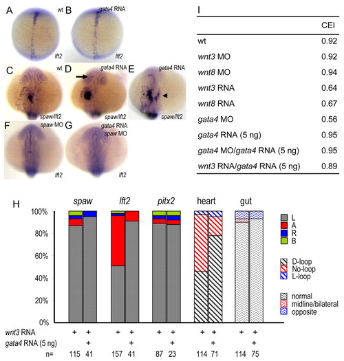
Gata4 mediates the function of Wnt signaling in regulating cardiac laterality. (A-G) Overexpression of gata4 induces lefty2 expression in a spaw-dependent manner. (A,B) Injection of gata4 RNA had no effect on lefty2 expression in floorplate precursors (B), compared with controls (A). Shown are dorsal views of 3-somite staged embryos with anterior to the top. (C-E) Injection of gata4 RNA induced lefty2 expression in diencephalon (D, arrow) and in the right heart field (E, arrowhead). (F,G) Injection of spaw morpholino resulted in a lack of lefty2 expression (F), which cannot be restored by gata4 RNA (G). Shown are dorsal views of 22-somite staged embryos with anterior to the top. (H) Overexpression of gata4 restored lefty2 expression and cardiac looping defects in embryos injected with wnt3 RNA. Shown is the quantification of side-specific gene expression, as well as heart and gut looping. L, left side; A, absence; R, right side; B, bilateral; n, total numbers of embryos scored. (I) Summary of CEI. Correlated expression between spaw and lefty2 was calculated by the co-efficiency index (CEI). Between 17 and 360 embryos were scored for each experimental group. EXPRESSION / LABELING:
|
|
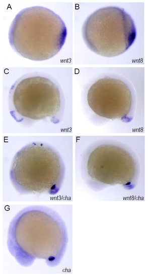
Wnt3 and Wnt8 are expressed in the vicinity of KV. (A-D) wnt3 and wnt8 expression was detected in the bud region of embryos at the bud stage (A,B) and the 12-somite stage (C,D). (E,F) The expression of wnt3 and wnt8 was in the vicinity of KV, as indicated by co-staining with charon. (G) Staining of charon alone at 12-somite staged embryos. Shown are lateral views with anterior to the left. EXPRESSION / LABELING:
|
|
The effect of Wnt/β-catenin signaling on cardiac jogging. (A-C) Representative images of cardiac left-jog (L-jog), No-jog and right-jog (R-jog). Shown are dorsal views of 28 hpf embryos after in situ hybridization using cmlc2 as a riboprobe. Anterior is to the top. (D) Quantification of embryos in each category of cardiac jogging. n, total numbers of embryos scored. |
|
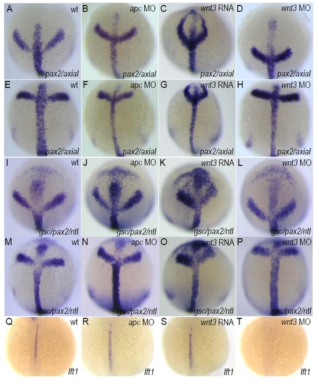
The effect of Wnt/β-catenin signaling on midline development. (A-P) Midline structures were not affected by alterations of Wnt signaling, as revealed by staining for the midline mesendodermal markers axial (A-H) or gsc and ntl (I-P). Embryos were co-stained with pax2 as an anteroposterior reference. Shown are dorsal views with anterior to the top of tailbud-staged embryos. (Q-T) lefty1 expression in the notochord was abolished in wnt3 morphants (T), but not in apc morphants (R) or in embryos injected with wnt3 RNA (S). Shown are dorsal views of 21-somite staged embryos with anterior to the top. EXPRESSION / LABELING:
|
|
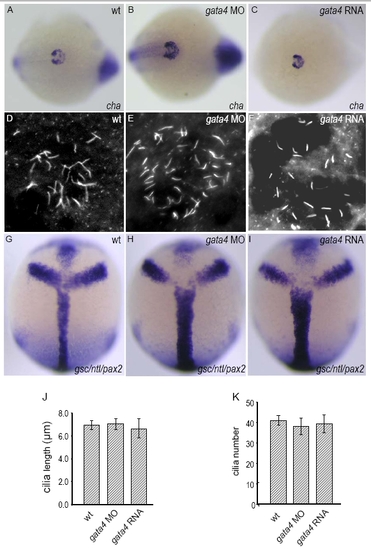
Gata4 is not required for KV development or midline development. (A-C) The expression of charon was not changed in gata4 morphants (B) or in embryos injected with gata4 RNA (C), when compared with controls (A) at the 12-somite stage. Shown are dorsal views of the tail bud region. (D-F) KV cilia development was not affected in gata4 morphants (E) or in embryos injected with gata4 RNA (F), when compared with controls (D) at the 12-somite stage, as visualized by anti-acetylated tubulin antibody staining; this is also summarized by measurement of cilia length (J) and cilia numbers (K). (G-I) Midline structure was not affected in gata4 morphants (H) or in embryos injected with gata4 RNA (I), as revealed by staining for the midline mesendodermal markers gsc and ntl. Shown are dorsal views of tailbud-staged embryos with anterior to the top. Data shown in J and K are means±s.d. Ten to 22 embryos were scored for each experimental group. EXPRESSION / LABELING:
|


