- Title
-
Bmp and Fgf signaling are essential for liver specification in zebrafish
- Authors
- Shin, D., Shin, C.H., Tucker, J., Ober, E.A., Rentzsch, F., Poss, K.D., Hammerschmidt, M., Mullins, M.C., and Stainier, D.Y.
- Source
- Full text @ Development
|
Bmp signaling is essential for hepatoblast specification. Embryos obtained from outcrossing a hemizygous Tg(hsp70l:dnBmpr-GFP) zebrafish were heat shocked at 18 hpf, and harvested at 29-32 (A-O) or 38-40 (P,Q) hpf. The expression of hhex (A-C), prox1 (D-F), gata4 (G-I), gata6 (J-L), foxa3 (M-O) and ceruloplasmin (cp) (P,Q) was then examined by in situ hybridization. The percentage of hemizygous embryos exhibiting a similar expression pattern is indicated in the lower left corner (n=8-10). (A-C) hhex expression in the liver region (arrows) was greatly reduced or almost absent in the hemizygous embryos, whereas its expression in the pancreatic islet (arrowheads) appeared unaffected. (D-F) prox1 expression in the liver region (arrows) was greatly reduced or almost absent in the hemizygous embryos, whereas its expression in the interrenal primordium (arrowheads) was less affected. To better visualize hepatic prox1 expression, a side-view image is shown in an inset. (G-L) gata4 and gata6 expression was reduced in the liver region (arrows), whereas their expression in the intestinal endoderm was barely affected (brackets). (M-O) The leftward bending of the gut, revealed by foxa3 expression, was often defective in the hemizygous embryos (brackets). (P,Q) Hepatocyte expression of cp was absent in the hemizygous embryos. All images, except insets, are dorsal views, anterior left. |
|
Alk8 is required for early liver development. Wild-type and alk8 mutant zebrafish embryos at 26 (A-H), 28 (I,J) and 48 (K,L) hpf were analyzed for hhex (A,B), prox1 (C,D), gata4 (E,F), gata6 (G,H), foxa3 (I,J) and cp (K,L) expression. (A,B) hhex expression in the liver region (arrows) is greatly reduced in the mutants, whereas its expression in the pancreatic islet appears unaffected (arrowheads). (C,D) prox1 expression in the liver region (arrows) is also greatly reduced in the mutants, whereas its expression in the interrenal primordium appears unaffected (arrowheads). To better visualize hepatic prox1 expression, a side-view image is shown in an inset. (E-H) gata4 and gata6 expression is reduced in the liver region (arrows), whereas their expression in the intestinal endoderm is only mildly affected (brackets). (I,J) The leftward bending of the gut appears unaffected in the mutants (brackets). (K,L) Hepatocyte expression of cp is completely absent in the mutants. All images, except insets, are dorsal views, anterior left. |
|
Bmp signaling is not essential for the maintenance of specified liver progenitors. Embryos obtained from outcrossing a hemizygous Tg(hsp70l:dnBmpr-GFP) zebrafish were heat shocked at 22 (A-C,G-I,M-O) or 26 (D-F,J-L,P-R) hpf, harvested at 33 (A-C,G-I), 36 (D-F,J-L) or 38-40 (M-R) hpf, and examined for hhex (A-F), prox1 (G-L) and cp (M-R) expression. The percentage of hemizygous embryos exhibiting a similar expression pattern is indicated in the lower left corner (n=8-10). When embryos were heat shocked at 22 hpf, both hhex and prox1 expression in the liver region (arrows) were greatly reduced in the hemizygous embryos, whereas their expression in other regions (arrowheads) appeared unaffected (B,C,H,I). By contrast, when they were heat shocked at 26 hpf, both hhex and prox1 were clearly expressed in the liver region in all the hemizygous embryos (E,F,K,L, arrows). However, hhex and prox1 expression in the liver region (arrows) was somewhat reduced compared with wild-type siblings, whereas hhex expression in the pancreatic islet and prox1 expression in the interrenal primordium (arrowheads) appeared unaffected (D-F,J-L). Hepatocyte differentiation, assessed by cp expression, barely occurred in the hemizygous embryos heat shocked at 22 hpf (N,O, arrows), and was clearly reduced in those heat shocked at 26 hpf (Q,R, arrows). All images, except insets, are dorsal views, anterior left. Insets are side views, anterior left. |
|
Overexpression of wild-type alk8 under a heat-shock promoter rescued liver defects in alk8 mutant zebrafish embryos. (A-R) Embryos obtained from crossing alk8+/- females with Tg(hsp70:alk8);alk8+/- males were heat shocked at 18 (A,C,G,I,M,O) or 34 (D,F,J,L,P,R) hpf, and harvested at 34 (A-C,G-I), 42 (D-F,J-L), 47 (M-O) or 54 (P-R) hpf. The expression of hhex (A-F), prox1 (G-L) and cp (M-R) was then examined (arrows). When alk8 was overexpressed at 18 hpf, the expression of hhex, prox and cp in the mutant embryos (C,I,O, arrows) was comparable to that in wild-type siblings (A,G,M, arrows), whereas their expression in the mutant embryos that were not heat shocked was strongly reduced (B,H,N, arrows). Even when alk8 was overexpressed at 34 hpf, hhex, prox and cp expression was substantial in the mutant embryos (F,L,R, arrows) compared with the mutant embryos that were not head shocked (E,K,Q, arrows). |
|
Gata4 and Gata6 are required for the expansion and differentiation of liver progenitors. Wild-type zebrafish embryos were injected with 10 ng gata4 MO and/or 2.5 ng gata6 MO, harvested at 37-38 hpf and examined for hhex (A-E), prox1 (F-J) and cp (K-N) expression. Injections of gata4 or gata6 MO weakly reduced hhex and prox1 expression in the liver region (B,C,G,H); injections of both MOs together strongly reduced their expression (D,E,I,J), but did not completely abolish it. Injections of gata4, gata6 or both MOs severely affected hepatocyte differentiation, as shown by cp expression (L-N). All images are dorsal views, anterior left (n=13-20). EXPRESSION / LABELING:
|
|
Fgf signaling is essential for hepatoblast specification. Embryos obtained from outcrossing a hemizygous Tg(hsp70l:dnfgfr1-EGFP) zebrafish were heat shocked at 18 hpf, harvested at 29-31 (A-N) or 38-40 (O,P) hpf, and examined for hhex (A-C), prox1 (D,E), gata4 (F-H), gata6 (I-K), foxa3 (L-N) and cp (O,P) expression. The percentage of hemizygous embryos exhibiting a similar expression pattern is indicated in the lower left corner (n=8-10). (A-C) hhex expression in the liver region (arrows) was greatly reduced or almost absent in the hemizygous embryos, whereas its expression in the pancreatic islet appeared unaffected (arrowheads). (D,E) prox1 expression in the liver region (arrows) was almost absent in the hemizygous embryos, whereas its expression in the interrenal primordium was less affected (arrowheads). To better visualize hepatic prox1 expression, a side-view image is shown in an inset. (F-K) gata4 and gata6 expression was greatly reduced in the liver region (arrows), whereas their expression in the intestinal endoderm was less affected (brackets). (L-N) The leftward bending of the gut did not occur in the hemizygous embryos (brackets). (P) Hepatocyte expression of cp was absent in the hemizygous embryos. All images, except insets, are dorsal views, anterior left. |
|
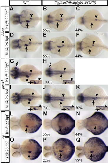
Fgf signaling is not essential for the maintenance of specified liver progenitors. Embryos obtained from outcrossing a hemizygous Tg(hsp70l:dnfgfr1-EGFP) zebrafish were heat shocked at 22 (A-C,G,H,L-N) or 26 (D-F,I-K,O-Q) hpf, harvested at 35-36 (A-K) or 38-40 (L-Q) hpf and examined for hhex (A-F), prox1 (G-K), and cp (L-Q) expression. The percentage of the hemizygous embryos exhibiting a similar expression pattern is indicated in the lower left corner (n=8-10). When embryos were heat shocked at 22 hpf, hhex expression in the liver region (arrows) was greatly reduced (B) or almost absent (C) in the hemizygous embryos, whereas its expression in the pancreatic islet (arrowheads) appeared unaffected (B,C). prox1 expression in the liver (black arrows) and retina (white arrows) was also greatly reduced in the hemizygous embryos (H), whereas its expression in the interrenal primordium appeared unaffected (H, arrowheads). By contrast, when embryos were heat shocked at 26 hpf, both hhex and prox1 were clearly expressed in the liver region in all the hemizygous embryos (E,F,J,K, arrows). However, hhex and prox1 expression in the liver region was reduced compared with wild-type siblings, whereas hhex expression in the pancreatic islet and prox1 expression in the interrenal primordium appeared unaffected (E,F,J,K, arrowheads). Hepatocyte differentiation, assessed by cp expression, was severely defective in the hemizygous embryos heat shocked at 22 hpf (M,N, arrows) and weakly reduced in those heat shocked at 26 hpf (P,Q, arrows). All images, except insets, are dorsal views, anterior left. Insets are side views, anterior left. |
|
Overexpression of Bmp2b under a heat-shock promoter partially rescued hepatoblast specification in zebrafish embryos lacking Fgf signaling. Embryos obtained from crossing a hemizygous Tg(hsp70:bmp2b)fr13 female with a hemizygous Tg(hsp70l:dnfgfr1-EGFP) male were heat shocked at 18 hpf and harvested at 30-32 (A-K) or 38-40 (L-Q) hpf. The expression of hhex (A-F), prox1 (G-K) and cp (L-Q) was then examined. hhex, prox and cp expression in the liver region mostly recovered in a majority of the embryos overexpressing Bmp2b and lacking Fgf signaling (E,F,J,K,P,Q, arrows), whereas their expression in embryos lacking Fgf signaling was strongly reduced (C,D,I,N,O, arrows). Twenty-five percent of the double hemizygous embryos did not show recovery of prox1 expression. The expression of hhex and prox1 in the embryos overexpressing Bmp2b (B,H, arrows) was comparable to that in wild-type siblings (A,G, arrows); cp expression was enhanced in the embryos overexpressing Bmp2b (M) compared with wild-type siblings (L). The percentage of the embryos exhibiting a similar expression is indicated in the lower left corner (n=11-12). All images are dorsal views, anterior up. |
|
Response of foregut endodermal cells to a transient block in Bmp signaling. (A-H) Embryos obtained from outcrossing a hemizygous Tg(hsp70l:dnBmpr-GFP) zebrafish were heat shocked at 18 hpf for 25 minutes, harvested at 2 (A,B), 3 (C-E) or 4 (F-H) dpf and examined for cp expression. Distinct, hepatocyte expression of cp in the heat-shocked hemizygous embryos was not detected at 2 dpf (B), but was detected in 20% of the embryos at 3 dpf and 60% at 4 dpf (E,H). The percentage of hemizygous embryos exhibiting a similar expression level is indicated in the lower left corner (n=9-11). Arrows point to the pectoral fins. (I) The percentage of embryos exhibiting distinct, hepatocyte cp expression in A-H together with that of embryos at 5 and 6 dpf is shown in the graph (n=9-11). Black circles and black squares denote wild-type siblings and hemizygous embryos, respectively. Embryos obtained from crossing a hemizygous Tg(hsp70l:dnBmpr-GFP) fish with a homozygous Tg(fabp1a:dsRed) fish were treated in the same way as above, and examined for DsRed expression under a dissecting fluorescence microscope. Distinct, DsRed expression in the hemizygous embryos was not detected by 4 dpf, but was detected in 33% of the embryos at 5 dpf (J-L). Red circles and red squares denote wild-type siblings and hemizygous embryos, respectively (n=20). Fluorescence (J), brightfield (K) and a merged (L) image of the embryos at 5 dpf are shown. Embryos transiently lacking Bmp signaling eventually initiated cp and fabp1a- DsRed expression, although with a delay. A-H, dorsal views, anterior left; J-L, ventrolateral views, anterior up. |
|
gata4 and gata6 are expressed in the endoderm at least from 18 hpf. Wild-type zebrafish embryos were harvested at 18 (A,E), 21 (B,F), 24 (C,G) and 26 (D,H) hpf and examined for gata4 (A-D) and gata6 (E-H) expression. Arrows point to the liver primordium. EXPRESSION / LABELING:
|
|
gata4 and gata6 expression in embryos in which Bmp signaling was blocked at 22 and 26 hpf. Embryos obtained from outcrossing a hemizygous Tg(hsp70l:dnBmpr-GFP) zebrafish were heat shocked at 22 (A-B,F-H) or 26 (C-E,I-K) hpf, harvested at 33 (A-B,F-H) or 36 (C-E,I-K) hpf, and examined for gata4 (A-E) and gata6 (F-K) expression. The first column shows wild-type siblings; the second and third columns show hemizygous embryos. The percentage of hemizygous embryos exhibiting a similar expression pattern is indicated in the lower left-hand corner (n=8-10). The expression of gata4 and gata6 in the liver region of the hemizygous embryos heat shocked at 22 hpf was reduced to a greater extent than that in the embryos heat shocked at 26 hpf. gata4 expression in the hemizygous embryos heat shocked at 26 hpf was still reduced compared with wild-type siblings, whereas gata6 expression in those embryos appeared barely affected (C-E versus I-K, arrows). Arrows point to the liver region. All images are dorsal views, anterior left. |
|
gata4 and gata6 expression in embryos in which Fgf signaling was blocked at 22 and 26 hpf. Embryos obtained from outcrossing a hemizygous Tg(hsp70l:dnfgfr1-EGFP) zebrafish were heat shocked at 22 (A-C,G-H) or 26 (D-F,I-K) hpf, harvested at 35-36 hpf, and examined for gata4 (A-E) and gata6 (F-K) expression. The percentage of hemizygous embryos exhibiting a similar expression pattern is indicated in the lower left-hand corner (n=8-10). (A-K) The expression of gata4 and gata6 in the liver region of the hemizygous embryos heat shocked at 22 hpf was similar to that in the embryos heat shocked at 26 hpf and only weakly reduced compared with wild-type siblings. Arrows point to the liver region. All images are dorsal views, anterior left. EXPRESSION / LABELING:
|


