
Fig. 2
|
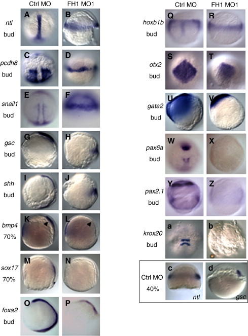
Fig. 2 Molecular marker expression in FoxH1-depleted embryos. Wild type embryos were injected with 8 ng of either control MO or FoxH1 MO1, fixed at the indicated stage and processed for whole-mount in situ hybridization. Probes, stages and MOs injected are as indicated. (A–J) Heterochronic expression patterns of mesoderm markers. Panels A–F are dorsal views, with the animal pole to the top; panels G–P are lateral views, with the dorsal side to the right. (K–P) Reduction of mesoderm and endoderm markers. Panels K–P are lateral views, with the dorsal side to the right. (Q–V) Persistence of broad neurectoderm markers in FoxH1 morphants. Panels Q, R are dorsal views, with the animal pole to the top; panels S, T are dorsal-animal views, with the animal pole somewhat below the top; panels U, V are lateral views, with the dorsal side to the right. (W–b) Disrupted neural patterning in FoxH1 morphants. Panels W–Z and a–b are dorsal views, with the animal pole to the top. (c–d) Expression of ntl and gsc in control embryos at the 40% epiboly stage. Panels c, d are lateral views, with the dorsal side to the right. Abbreviations and stages: bud, bud stage (10 hpf); 70%, 70% epiboly stage (8 hpf); 40%, 40% epiboly stage (5 hpf); FH1, FoxH1; ctrl, control.
Reprinted from Developmental Biology, 310(1), Pei, W., Noushmehr, H., Costa, J., Ouspenskaia, M.V., Elkahloun, A.G., and Feldman, B., An early requirement for maternal FoxH1 during zebrafish gastrulation, 10-22, Copyright (2007) with permission from Elsevier. Full text @ Dev. Biol.