
Fig. 9
- ID
- ZDB-FIG-250430-202
- Publication
- Hammond et al., 2025 - Modularity of the segmentation clock and morphogenesis
- Other Figures
-
- Fig. 1
- Fig. 2
- Fig. 2 - Supplemental 1
- Fig. 3
- Fig. 3 - Supplemental 1
- Fig. 4
- Fig. 4 - Supplemental 1
- Fig. 4 - Supplemental 2
- Fig. 4 - Supplemental 3
- Fig. 4 - Supplemental 4
- Fig. 4 - Supplemental 5
- Fig. 5
- Fig. 5 - Supplemental 1
- Fig. 5 - Supplemental 2
- Fig. 5 - Supplemental 3
- Fig. 6
- Fig. 6 - Supplemental 1
- Fig. 6 - Supplemental 2
- Fig. 6 - Supplemental 3
- Fig. 7
- Fig. 7 - Supplemental 1
- Fig. 8
- Fig. 8 - Supplemental 1
- Fig. 8 - Supplemental 2
- Fig. 8 - Supplemental 3
- Fig. 9
- Fig. 10
- All Figure Page
- Back to All Figure Page
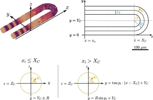
Geometry of the pre-somitic mesoderm (PSM) assumed in the present model. Top left: Major axes (x , y , z ) used in the model. x corresponds to the anterior-posterior axis of the embryo and increases towards the tissue posterior, y corresponds to the left-right axis and increases to the right-hand side of the tissue, and z corresponds to the dorsal-ventral axis of the embryo. Top right: Schematic of the PSM in the xy plane. The PSM is comprised of two cylinders, centred at y=rT and y=2R+rT , respectively, with radius rT . The ‘tailbud’ is represented as a half-torus subdomain centred at x=(Xc,Yc,Zc)T , with minor radius rT and major radius R . Bottom: Cross sections of the tissue showing how a point xi is assigned the polar coordinates ri and qi , in both the PSM cylinders (xi≤Xc ) and the half-toroid tailbud (xi>Xc ). Adapted from Uriu et al., 2021. |