|
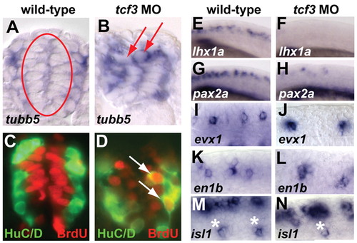
Spinal progenitors lacking Tcf3 precociously express neuronal markers. (A,B) Cross-sections of tubb5 in situ hybridization at 18 hpf show that tcf3 morphants have ectopic tubb5-positive cells in the medial progenitor domain (arrows). (C,D) Double labeling for HuC/D (green) and BrdU (red) at 24 hpf shows BrdU-positive Hu-positive cells (arrows) in tcf3 morphants, some of which are in the medial domain. (E-N) Lateral whole-mount views of postmitotic neuronal markers at 18 hpf. (E-H) lhx1a and pax2a, which mark multiple classes of spinal interneurons, are drastically reduced in tcf3 morphants. (I-N) evx1+ interneurons, en1b+ interneurons and isl1+ primary motoneurons (asterisks) are also reduced in tcf3 morphants.
|