
- Title
-
Thyroid development in zebrafish lacking Taz
- Authors
- Pappalardo, A., Porreca, I., Caputi, L., De Felice, E., Schulte-Merker, S., Zannini, M., Sordino, P.
- Source
- Full text @ Mech. Dev.
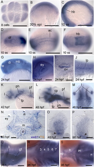
Zebrafish wwtr1 mRNA expression. WISH data from 8-cell stage to 96 hpf. Wwtr1 mRNA is maternally provided (A) and then ubiquitously distributed at low levels during gastrulation (B) and early somitogenesis (C), when a strong signal is detected in dorsal forebrain (D), hindbrain (E) and adaxial cells (F). Diffuse expression at 24 hpf, with strong expression in brain (G), eye (lens and retina) (H), otic vesicles (I) and lateral line primordium (J). At 42 and 48 hpf, wwtr1 mRNA is seen in lens, pectoral fins and pharyngeal area (K–N). Transverse section of a 60 hpf embryo showing a large domain of wwtr1 mRNA-positive cells covering the pharyngeal tissue and the thyroid follicle with TG immunostaining depicted (N). At 96 hpf, wwtr1 is expressed in pectoral fins (P), liver and gut (Q), anterior craniofacial (R) and neurocranial (S) cartilages. Arrowheads indicate higher level of expression in the pectoral fin mesenchyme proximally (O and P) Scale bar = 200 µm. 3–7, (branchial) aortic arch arteries; df, dorsal forebrain, ac, adaxial cells; cp, cephalic floor plate; di, diencephalon; ep, ethmoid plate; ey, eye; fm, fin mesenchyme; ff, fin fold; gt, gut; hb, hindbrain; le, lens; l, liver; lp, lateral line primordium; m, mouth; mb, midbrain-hindbrain boundary; n, notochord; ov, otic vesicle; pc, polar cartilage; pf, pectoral fin; ph, pharyngeal tissue; te, telencephalon; tl, thyroid follicle lumen; tf, thyroid follicular cells; tp, thyroid primordium; tr, trabeculae. Anterior is to the left in dorsal (A, D–F), ventral (R and S) and lateral view (B, C, G–L, Q). In (M), anterior is to the top in dorsal view. |

wwtr1-MO injection effect on the expression of thyroid transcription factors and thyroid specific genes. 48 hpf zebrafish embryos uninjected (uninj) (A and C) and injected with standard control-MO (scMO) (E, G, I) and wwtr1-MO (B, D, F, H, J), hybridized with probes for thyroid transcription factors nkx2.1a, pax2a and pax8, and thyroid differentiation genes tg and slc5a5 (A–H). Arrowheads indicate the thyroid gland. Asterisk depicts specific nkx2.1a mRNA signal over high staining background (B). Scale bar = 200 µm. Anterior is to the left in lateral view. |

ZFIN is incorporating published figure images and captions as part of an ongoing project. Figures from some publications have not yet been curated, or are not available for display because of copyright restrictions. EXPRESSION / LABELING:
PHENOTYPE:
|

Whole-mount immunostaining of T4 and TG. Uninjected (Uninj) (A), injected with standard control-MO (scMO) (C and E) and wwtr1-MO (B, D, F) 4 dpf larvae were immunolabeled for T4 (A-D) and TG (E and F). Thyroid follicles are reduced in number (A-D) and not restricted to the ventral midline in Taz-deprived larvae (C-F). Arrowheads indicate thyroid follicles. Scale bar represents 200 µm (A, B, E, F) and 100 µm (C and D). Anterior is to the left in ventral (A-D) and lateral views (E and F). |
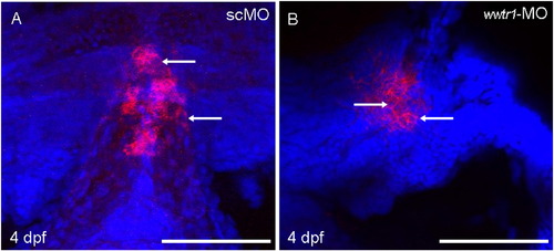
Zebrafish Taz controls the number of thyroid follicular cells. Z-stack images of tg mRNA labeling in DAPI stained zebrafish at 4 dpf injected with standard control-MO (A) and wwtr1-MO (B). Arrows indicate nuclei of tg expressing thyroid follicle cells. Scale bar represents 100 µm. PHENOTYPE:
|
|
|
ZFIN is incorporating published figure images and captions as part of an ongoing project. Figures from some publications have not yet been curated, or are not available for display because of copyright restrictions. PHENOTYPE:
|
Reprinted from Mechanisms of Development, 138 Pt 3, Pappalardo, A., Porreca, I., Caputi, L., De Felice, E., Schulte-Merker, S., Zannini, M., Sordino, P., Thyroid development in zebrafish lacking Taz, 268-78, Copyright (2015) with permission from Elsevier. Full text @ Mech. Dev.