
- Title
-
Ptk7 promotes non-canonical Wnt/PCP-mediated morphogenesis and inhibits Wnt/beta-catenin-dependent cell fate decisions during vertebrate development
- Authors
- Hayes, M., Naito, M., Daulat, A., Angers, S., and Ciruna, B.
- Source
- Full text @ Development
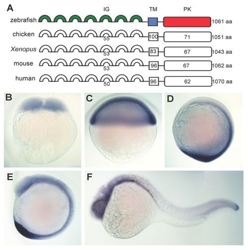
Zebrafish Ptk7 structure and embryonic expression. (A) Domain structure of zebrafish Ptk7 and its homology to chicken (KLG), Xenopus, mouse and human orthologues. Numbers indicate percentage amino-acid identity within extracellular immunoglobulin (IG), transmembrane (TM) and intracellular pseudokinase (PK) domains. (B-F) WISH of ptk7 expression throughout the first 24 hours of development. Lateral views of two-cell (0.75 hpf, B), shield (6 hpf; C), bud (10 hpf; D), 10- to 12-somite (15 hpf; E) and 24 hpf (F) stage embryos are shown. |
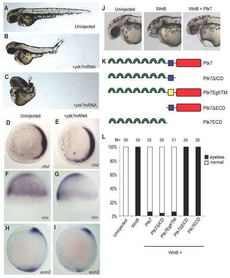
Ptk7 overexpression disrupts embryonic patterning and morphogenesis and inhibits exogenous Wnt/β-catenin activity. (A) Lateral view of uninjected control embryos at 36 hpf. (B,C) Embryos injected with ptk7 (400 pg) mRNA at the one-cell stage exhibit axial morphogenesis (B) and dorsoventral patterning defects (C). (D,E) Whole-mount in situ hybridisation (WISH) demonstrating chordin (chd) expression in control (D) and ptk7 (400 pg) mRNA-injected (E) embryos at shield stage (animal pole view, dorsal right). Arrowhead indicates lateral expansion of chd expression in embryos overexpressing Ptk7. (F,G) WISH of vox expression in control (F) and ptk7 (400 pg) mRNA-injected (G) embryos at shield stage (lateral view, dorsal right). (H,I) WISH for axin2 in control (H) and ptk7 (400 pg) mRNA-injected (I) embryos at bud stage (lateral view). Reduced vox and axin2 expression is observed in embryos overexpressing Ptk7. (J) Overexpression of wnt8 (10 pg) disrupts CNS pattern, as demonstrated by loss of eyes and reduced forebrain at 36 hpf. Co-injection of ptk7 (300 pg) mRNA can largely rescue these wnt8-induced phenotypes. (K) Schematic of Ptk7 mutant isoforms generated for structure-function and rescue experiments. (L) Quantification of phenotypes observed upon injection of wnt8 (10 pg) mRNA, and upon co-injection of wnt8 with full length ptk7 (300 pg), ptk7ΔICD (200 pg), ptk7 egfrTM (300 pg), ptk7ΔECD (150 pg) and ptk7 ECD (200 pg) mRNA. mRNA concentrations were adjusted to yield equimolar amounts of truncated/mutant Ptk7 protein. Embryos were scored as being ‘eyeless’ if the eye was either completely absent or <25% the size of uninjected controls. |

Ptk7hsc9 mutant transcript is targeted for non-sense-mediated decay (NMD). (A) Ptk7 ZFN target sequence and ptk7hsc9 mutant allele. Both the nucleotide and protein sequence are represented. Ptk7 ZFNs induced a 10-bp deletion, represented by dashed lines in the mutant sequence. This frame-shift mutation yields a premature termination codon immediately adjacent to the target site. Boxes outline the nucleotide target of left (ZFN L) and right (ZFN R) zinc-finger proteins. SfaN1 indicates the restriction site used to identify potential mutations. (B) Mutations were identified by PCR amplification of genomic DNA followed by SfaN1 restriction digest. Examples of wild-type (+/+), heterozygote (+/-) and mutant (-/-) zebrafish are represented. The Ptk7hsc9 allele is not targeted by SfaN1, and runs higher on a DNA gel than wild-type ptk7. (C,D) Wild-type (C) and zygotic ptk7hsc9 mutant (D) zebrafish at 2 months post-fertilisation. Axial curvatures were observed in 100% of ptk7hsc9 mutant juvenile and adult zebrafish. (E-I) ptk7 expression in wild-type (E,G) and maternal-zygotic ptk7hsc9 (MZptk7hsc9) mutant (F,H,I) embryos, as visualised by WISH at the one-cell (E,F), 10- to 12-somite (G,H) and 24 hpf (I) stages. (J-L) Quantitative RT-PCR (qRT-PCR) reveals a strong reduction in ptk7 transcript in MZptk7hsc9 relative to wild-type at shield (J; ***P<0.001), bud (K; ***P<0.001) and 10- to 12-somite (L; *P=0.0358) stages. Error bars represent the s.e. for the expression level fold change. PHENOTYPE:
|
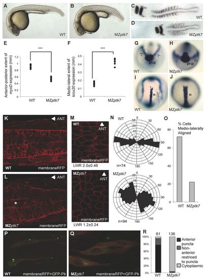
MZptk7 mutant embryos display PCP-mediated morphogenesis defects. (A,B) Lateral views of wild-type (A) and MZptk7hsc9 mutant (B) embryos at 24 hpf. (C,D) Flat-mounts of 10- to12 somite-stage wild-type (C) and MZptk7hsc9 (D) embryos stained for krox20 (hindbrain) and myoD (somite) gene expression. (E) Quantification of the anterior-posterior extent of the myoD expression domain in wild-type (WT) versus MZptk7hsc9 mutant embryos (***P<0.001, n=8 for each group). (F) Quantification of the mediolateral extent of krox20 expression in WT versus MZptk7hsc9 mutant embryos (***P<0.001, n=8 for each group). MZptk7hsc9 mutant embryos display clear defects in axial extension. (G-J) Anterior (G,H) and dorsal (I,J) views of bud stage WT (G,I) and MZptk7hsc9 (H,J) embryos stained for hgg1 (prechordal plate, pcp), dlx3 (prospective neural plate, arrowheads) and ntl (prospective notochord, n). MZptk7hsc9 mutants demonstrate defects in the convergence of both neuroectoderm and axial mesoderm tissues. (K,L) Dorsal confocal images of the neural tube and adjacent somites of 24 hpf wild-type (K) and MZptk7hsc9 (L) embryos, injected with membrane-localised monomeric RFP (membraneRFP) (Megason and Fraser, 2003). MZptk7hsc9 mutant embryos display an accumulation of neural progenitors (asterisk in L) at the centre of the neural primordium. Anterior is left. (M) MembraneRFP-labelled cells in the dorsal ectoderm of WT and MZptk7hsc9 embryos at 90% epiboly. Dorsal view, midline to the right and anterior to the top. The length-to-width (LWR) ratio of cells are as indicated for WT (n=74) and MZptk7hsc9 (n=94). (N) Rose diagrams for cell orientation relative to the embryonic midline at 90% epiboly in WT and MZptk7hsc9 embryos. (O) Graph showing percentage of mediolaterally aligned cells, for which longitudinal axis is oriented ±15° with respect to the embryonic mediolateral axis. (P,Q) Dorsal confocal images of the neural keel and adjacent somites of 8- to 10-somite-stage WT (P) and MZptk7hsc9 (Q) embryos scatter-labelled with GFP-Prickle (GFP-Pk) and membrane RFP. The subcellular localisation of the PCP marker GFP-Pk is disrupted in MZptk7hsc9. Anterior is up. Confocal imaging was carried out at the level of the first to the fifth somite pairs. (R) Quantification of the localisation of GFP-Pk puncta in WT (four embryos) versus MZptk7hsc9 (six embryos). PHENOTYPE:
|
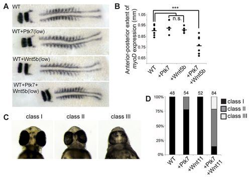
Ptk7 overexpression inhibits exogenous non-canonical Wnt/PCP activity. (A) Flat-mounts of 10- to 12-somite stage wild-type (WT) embryos and embryos injected with ptk7 (200 pg), wnt5b (50 pg) or ptk7 (200pg) + wnt5b (50 pg) mRNA stained for krox20 (hindbrain) and myoD (somite) gene expression. (B) Quantification of the anterior-posterior extent of the myoD expression domain in WT, ptk7 (200 pg), wnt5b (50 pg), and ptk7 (200 pg) + wnt5b (50 pg) mRNA-injected embryos (***P<0.001). n.s., not significant. (C) Ventral views of embryos at 2 days post-fertilisation (dpf). Cyclopia phenotypes were observed as class I (no cyclopia), class II (partial cyclopia) and class III (full cyclopia). (D) Quantification of cyclopia phenotypes in WT, ptk7 (200 pg), wnt11 (50 pg), and ptk7 (200 pg) + wnt11 (50 pg) mRNA-injected embryos at 2 dpf. |
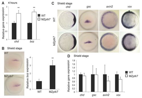
MZptk7hsc9 embryos establish normal dorsoventral pattern. (A) Chordin (chd) and bozozok (boz) expression is slightly increased in MZptk7hsc9 embryos immediately following MBT, as assayed by qRT-PCR of embryos at 4 hpf. **P<0.01. (B) Boz expression is increased in MZptk7hsc9 embryos prior to gastrulation as assayed by WISH (dorsal view), and by qRT-PCR at 50% epiboly (**P=0.0078). (C) Whole-mount in situ hybridisation (WISH) for dorsal organiser genes chordin (chd) and goosecoid (gsc), and ventral genes axin2 and vox at shield stage. Embryos are viewed from the animal pole with dorsal to the right (for chd, axin2 and vox). gsc expression is shown as a dorsal view. (D) qRT-PCR assays of chd (P=0.4675), gsc (P=0.9515), axin2 (P=0.07) and vox (P=0.3974) expression in wild-type (WT) versus MZptk7hsc9 embryos at shield stage. No significant differences in gene expression were observed. Error bars represent the s.e. for the expression level fold change. |

MZptk7hsc9 embryos establish normal dorsoventral pattern. (A) Chordin (chd) and bozozok (boz) expression is slightly increased in MZptk7hsc9 embryos immediately following MBT, as assayed by qRT-PCR of embryos at 4 hpf. **P<0.01. (B) Boz expression is increased in MZptk7hsc9 embryos prior to gastrulation as assayed by WISH (dorsal view), and by qRT-PCR at 50% epiboly (**P=0.0078). (C) Whole-mount in situ hybridisation (WISH) for dorsal organiser genes chordin (chd) and goosecoid (gsc), and ventral genes axin2 and vox at shield stage. Embryos are viewed from the animal pole with dorsal to the right (for chd, axin2 and vox). gsc expression is shown as a dorsal view. (D) qRT-PCR assays of chd (P=0.4675), gsc (P=0.9515), axin2 (P=0.07) and vox (P=0.3974) expression in wild-type (WT) versus MZptk7hsc9 embryos at shield stage. No significant differences in gene expression were observed. Error bars represent the s.e. for the expression level fold change. EXPRESSION / LABELING:
PHENOTYPE:
|
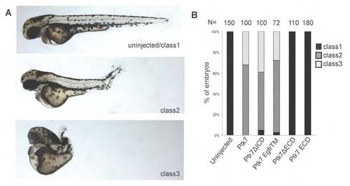
Plasma membrane-tethered extracellular domain is required for Ptk7 overexpression activity. (A) Class of phenotypes induced by ptk7 overexpression: wild-type/class 1; class 2, axial extension defects as well as dorsal curvatures of the posterior tail; class 3, mild to severe dorsalisation. (B) Distribution of phenotypes in embryos injected with ptk7 (400 pg), ptk7ΔICD (300 pg), ptk7 egfrTM (400 pg), ptk7ΔECD (300 pg) and ptk7 ECD (300 pg) mRNA. |
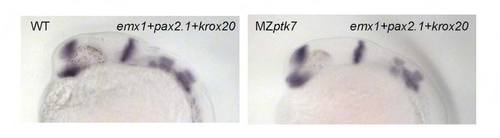
MZptk7hsc9 embryos do not display CNS dorsoventral patterning defects. Whole-mount in situ hybridization of wild-type (WT) and MZptk7 embryos at 24 hpf stained for emx1, pax2.1 and krox20 to mark the forebrain, midbrain-hindbrain boundary and posterior rhombomeres, respectively. PHENOTYPE:
|
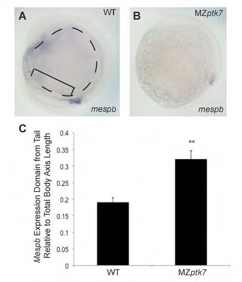
MZptk7hsc9 mutants display tailbud patterning defects. (A,B) Whole-mount in situ hybridization of mespb expression, which delineates the anterior extent of pre-somitic mesoderm in wild-type (A) and MZptk7hsc9 mutant (B) embryos. Mespb expression is shifted anterior relative to total body axis length in MZptk7hsc9. (C) Quantification of the ratio of the distance between the most posterior part of the embryonic tail (bracket in A) and the most posterior extent of the mespb expression domain to the total body axis length (dashed line in A) from head to tail in wild-type (WT) and MZptk7hsc9 at the 10- to 12-somite stage. Student’s t-test, **P=0.0016. Error bars represent s.d. EXPRESSION / LABELING:
|
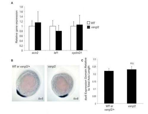
Abnormal PCP does not disrupt Wnt/β-catenin signaling or posterior tissue fate specification. (A) qRT-PCR to analyze expression of Wnt/β-catenin target genes in vangl2 mutants relative to wild-type (WT) reveals no difference in expression levels of axin2 (P=0.6314, Student’s t-test), lef1 (P=0.3469, Student’s t-test), or cyclin D1 (P=0.8825, Student’s t-test). Error bars represent standard error of the fold change. Each graph is representative of two independent experiments with three technical replicates each. (B) Lateral views of whole-mount in situ hybridization for the posterior mesodermal marker tbx6 in wild-type/vangl2+/– and vangl2 mutant embryos. (C) Quantification of the size of the tbx6 expression domain relative to the total embryonic body length in WT and vangl2 mutant embryos. The tbx6 expression domain is not significantly expanded in vangl2 mutant embryos. n.s., not significant, Students t-test. |