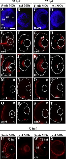
Morpholino-mediated depletion of rx1 results in lamination defects and reduced retinal cell differentiation. Embryos were treated at the 1–2 cell stage with a morpholino cocktail containing ATG (translational start site, 100 μM) and GT (splice donor site, 50 μM) directed morpholinos. (A–D) 5 μm sections stained with DAPI obtained at 53 hpf (A, B) and 72 hpf (C, D) from control (A, C) and rx-1-depleted (B, D) embryos, showing inhibition/delay of lamination in treated retinas. (E–H) Sectioned retinas stained for the ganglion cell surface marker, zn-8, evaluated at 53 hpf (E, F) and 72 hpf (G, H) from control (E, G) and rx1-depleted (F, H) retinas, showing patchy staining of the gcl as a consequence of treatment. (I–L) Sections stained for the amacrine and ganglion cell marker HuC/D, obtained at 53 hpf (I, J) and 72 hpf (K, L) from control (I, K) and rx1-depleted (J, L) embryos, showing reduced staining as a consequence of treatment. (M–P) Sections stained with the red/green double cone marker zpr1, from embryos fixed at 53 hpf (M, N) and 72 hpf (O, P) following treatment with control (M, O) and rx1-depleting (N, P) morpholinos, showing reduced cone differentiation in the treated retinas. (Q–T) Sections stained with the rod marker zpr3, from embryos fixed at 53 hpf (Q, R) and 72 hpf (S, T) following treatment with control (Q, S) and rx1-depleting (R, T) morpholinos, showing reduced rod differentiation in the treated retinas. (U–X) Sections stained for the rod bipolar cell marker, PCK (U, V) and the Müller glia cell marker GS (W, X) from control (U, W) and rx1-depleted (V, X) embryos fixed at 72 hpf, showing reduced differentiation of rod bipolar cells and the absence of Müller glia in treated retinas. le = lens; gcl = ganglion cell layer; inl = inner nuclear layer; onl = outer nuclear layer; V = ventral; D = dorsal; dotted lines indicate retinal boundary; white circles depict the location of the lens; scale bar = 50 μm.
|
