FIGURE SUMMARY
Title

Yap-lin28a axis targets let7-Wnt pathway to restore progenitors for initiating regeneration

Authors
Ye, Z., Su, Z., Xie, S., Liu, Y., Wang, Y., Xu, X., Zheng, Y., Zhao, M., Jiang, L.
Source
Full text @ Elife

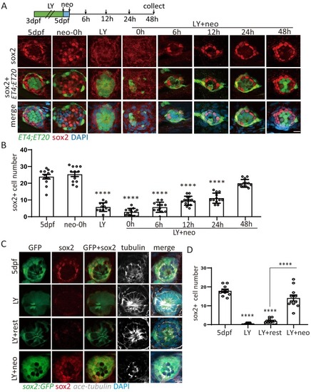
(A, B) ET4;ET20 larvae were treated with neomycin, LY411575 (3dpf-5dpf), or neomycin following LY (LY+neo), and collected at indicated time points post neomycin treatment for sox2 immunostaining. The number of sox2+ progenitors was not affected post neo, while it was significantly decreased in LY and LY+neo-0h. The sox2+ progenitors were regenerated post LY+neo and recovered to normal level at 48 hr post LY+neo. (C, D) The sox2:GFP reporter was treated with LY from 3dpf to 5dpf to exhaust GFP+ progenitors. GFP+ progenitors cannot be regenerated when resting in normal medium for 2 days post LY treatment (LY+rest). In contrast, sox2+ progenitors were quickly recovered to normal level at 2-day post LY+neo. Scale bar equals 10 μm. All groups are compared with 5dpf unless indicated

Severe injury causes damage to HCs, SCs and MCs.

ET4;ET20 larvae were treated with 2 μM LY from 3dpf to 5dpf followed by neomycin, and the number of HCs, SCs and MCs were counted before and right after neo treatment. Results showed that HCs were increased while SCs were decreased post LY, indicating that LY induced differentiation of SCs into HCs. Numbers of HCs, SCs and MCs were all significantly decreased post LY+neo compared with 5dpf normal larvae. Scale bar equals 10 μm.

More proliferative SCs and MCs were induced post severe injury compared with normal injury.

Larvae treated with neo or LY+neo were incorporated with EdU for different time points and counted for numbers of differentiating cells (EdU+ET4+ cells, HC+), proliferative SCs (EdU+ET4-ET20- cells, SC+), and proliferative MCs (EdU+ET20+ cells, MC+). We calculated EdU incorporation per hour by dividing EdU+ cell number with incorporation time. Results showed that differentiation rate (HC+ in B) in LY+neo was decreased compared with neo, while proliferative rates of SCs (SC+ in C) and MCs (MC+ in D) were significantly increased. Scale bar equals 10 μm.

<italic>lin28a</italic> was induced by various kinds of injury and <italic>lin28a</italic> expression level was proportional to injury size.

(A) RNA-Seq analysis using neomycin-treated neuromast cells revealed that lin28a was highly induced at 1 hr post neo while lin28b expression wa s not changed. (B) lin28a was induced at 1 hr post treatments with neomycin, cisplatin and copper. (C) LY-treated larvae were subjected for laser scan to ablate HCs at the indicated locations (white circles). Expression levels of lin28a was proportional to the injured cell number manipulated by laser ablation. (D) Laser was targeted at the circles for ablating indicated number of HCs or SCs. The corresponding neuromast was performed for lin28a in situ, and the results showed that either HC damage or SC damage could induce lin28a expression. Scale bar equals 10 μm.

<italic>Yap</italic> was activated immediately post LY+neo.

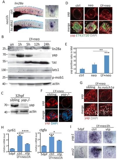
(A) Wnt inhibition with hs:dkk1 has no effect on lin28a induction post neo treatment. (B) Expression levels of lin28a, yap, taz and Hippo pathway genes, such as lats1 and mob1, were detected by western blot at various time points post LY+neo. Results showed that yap was increased at 1 hr post LY+neo. Levels of lats1 and phosphorylated mob1 were not changed at 1 hr, suggesting yap is activated independent of canonical Hippo pathway. (C) Specificity of yap antibody was tested with 32hpf sibling and yap mutants. (D–E) Larvae treated with neo or LY+neo were immonstained with yap antibody. Results showed that nuclear localized yap was increased in LY+neo compared with neo. (F) LY+neo induced yap expression in sibling but not yap mutants. (G) Yap activation post LY+neo was inhibited when atoh1a is inhibited with hs:notch1a, suggesting that yap is activated in atoh1a+ HC precursors. (H) Classic yap target genes cyr61 and ctgfa were upregulated post LY+neo, which is partially blocked by verteporfin. (I) Transcripts of yap and mst2 were highly induced post LY+neo, which was blocked by verteporfin. Scale bar equals 10 μm.

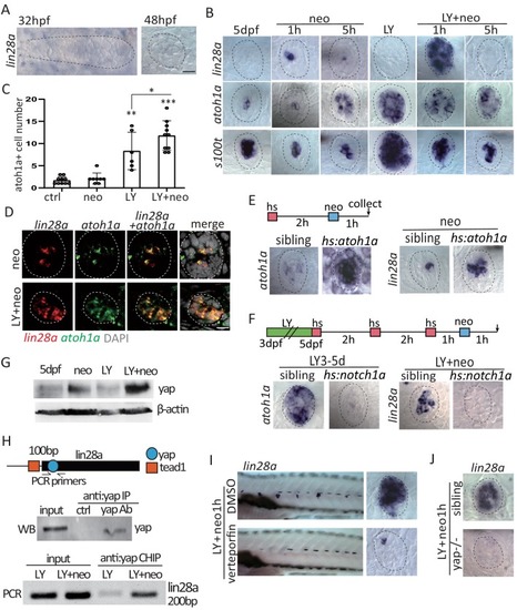
(A) Lin28a was not expressed in the developing lateral line primordium or neuromast. (B) Treatment with Notch inhibitor LY411575 from 3dpf to 5dpf increased expression atoh1a and s100t. More lin28a expression was observed in LY+neo-1h compared with neo-1h. No lin28a was detected at 5 hr post LY+neo or neo. (C) The number of atoh1a-transcribed cells detected by in situ were higher at 1 hr post LY+neo compared with LY. (D) Double fluorescent in situ showed that lin28a was co-expressed with atoh1a post neo or LY+neo. (E) Induction of lin28a post injury was increased when atoh1a was overexpressed in hs:atoh1a. (F) Lin28a expression was completely blocked when atoh1a was inhibited in hs:notch1a. (G) Western blot results showed that LY+neo induced higher yap expression compared with neo alone. (H) Motifs of yap and tead1 (co-transcriptional factor of yap) binding sites were predicted near lin28a transcriptional start site. CHIP-PCR results verified that yap directly binds the predicted motif. (I and J) Inhibition of yap using verteporfin or yap mutant blocked lin28a induction post LY+neo. Scale bar equals 10 μm.

Create <italic>lin28a</italic> mutant using CRISPR.

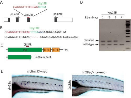
(A) Diagram shows four exons of lin28a and the position of CIRPSR that was used for creating lin28a mutant. (B–C) Deletion of five nucleotides in the lin28a mutant allele damages the restriction enzyme site of Hpy188I and leads to a truncated protein by creating a premature stop codon in lin28a mRNA. (D) F1 embryos were genotyped for lin28a mutation by PCR followed by Hyp188I digestion. (E) Transcribed lin28a mRNA level was decreased in lin28a-/- post LY+neo, probably because the mutant form of lin28a mRNA is unstable and degraded.

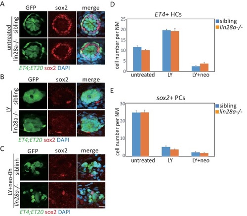
HCs and sox2<sup>+</sup> progenitors were not affected by <italic>lin28a</italic> deficiency in homeostasis, LY and LY+neo.

We compared numbers of ET4+ HCs and sox2+ progenitors in 5dpf sibling and lin28a-/- and found that they were not changed in lin28-/- in homeostasis (A), LY (B) and LY+neo (C). Scale bar equals 10 μm.

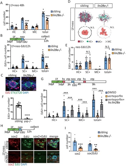
(A) The number of SCs and total cells were counted at 48 hr post LY+neo and were significantly decreased in lin28a mutant compared with sibling. (B–E) The ET4;ET20 larvae were incorporated with EdU post neo or LY+neo treatment. Three populations labeled with ET4+EdU+ (HC+), ET20+EdU+ (MC+) or ET4-ET20-EdU+ (SC+) were counted and recorded with location information. The proliferative SCs post LY+neo were significantly decreased in lin28a mutant compared with sibling (B and C), while not changed in neo-induced regeneration (E). (D) EdU plots show the positions of EdU+ nuclei of 18 neuromasts superimposed on the same plane, and rose diagrams document the angular positions of SC+. The results show that the proliferative SCs are evenly distributed in each quadrant with no polarization post LY+neo. (F) LY+neo-induced Edu incorporation was significantly reduced in yap mutant. (G) The hs:lin28a larvae were heat-shocked and pre-treated with 10 μM verteporfin before adding neomycin. Samples were collected at 14 hr post LY+neo for EdU+ cell counting. Proliferation (SC+) is significantly decreased post LY+neo in verteporfin, which could be rescued by overexpression of lin28a with hs:lin28a. (H, I) The numbers of regenerated sox2+ progenitors (sox2) and proliferative progenitors (sox2EdU) post LY+neo were both reced in lin28a mutant. Scale bar equals 10 μm.

(A–D) ET4;ET20;hs:lin28a larvae were incorporated with BrdU for 24 hr following heat-shock and/or neomycin treatment. (A, B) Overexpression of lin28a increased number of proliferative SCs (SC+) in both untreated and neomycin conditions. (C, D) BrdU plots and rose diagrams indicate that locations of SC+ still remain dorsally and ventrally polarized in hs:lin28a. (E, F) OverexpressUion of lin28a is sufficient to partially restore the exhausted sox2+ progenitors post LY. Scale bar equals 10 μm.

Developing HCs and MCs were not affected in hs:lin28a.

The ET4;ET20;hs:lin28a larvae were heat-shocked at 3dpf or 5dpf and collected after 24 hr for HC and MC counting. Results showed that numbers of HCs and MCs were not significantly changed by overexpression of lin28a at 3dpf (A) and 5dpf (B and C).

(A–B) Larvae were stained with sox2 antibody and atoh1a RNA probe at 6 hr post neo or LY+neo treatment. The number of atoh1a+sox2+ cells was significantly increased in LY+neo group. (C–D) The atoh1a:TdTomato larvae were used to trace atoh1a+ HC precursors in ctrl, LY or LY+neo. Results showed that Tomato+ cells labeled partial ET4+ HCs and ET4-sox2- HC precursors (yellow arrows). However, many atoh1a+ cells start to express sox2 from 12 hr post LY+neo and their numbers were significantly increased compared with normal larvae. The arrowheads in (C) pointed the atoh1a+sox2+ cells at 48 hr post LY+neo. (E) The atoh1a:TdTomato;hs:lin28a larvae was treated with LY+neo and verteporfin and collected for immunostaining with sox2 antibody. The cell number of Tomato+sox2+ is decreased in verteporfin and overexpression of lin28a could rescue the phenotype. (F–G) The LY-treated hs:lin28a larvae were heat-shocked to overexpress lin28a. Samples were collected for staining with sox2 antibody and atoh1a RNA probe. The number of atoh1a+sox2+ cells was significantly increased in hs:lin28a group, indicating that lin28a is sufficient to express sox2 in atoh1a+ HC precursors. Scale bar equals 10 μm.

Atoh1a+ cells became SCs and MCs post severe injury.

(A) The Neo-treated atoh1a:TdTomato;brn3c:GFP larvae was imaged for time lapse. Results showed that one Tomato+ cell turned into two HCs with membrane-bound GFP post neo (blue dots). (B) The LY+neo-treated atoh1a:TdTomato;sox2:GFP larvae was imaged for time lapse. The blue dots indicate that one Tomato+ cell divided and turned into two sox2:GFP-negative HCs post LY+neo. The white dots indicate that two Tomato+ cells divided and became four sox2:GFP-positive cells post LY+neo. (C and D) The atoh1a:TdTomato;ET4;ET20 larvae were collected at 48 hr post neo or LY+neo. Tomato expressing HCs, SCs and MCs were separately divided by total numbers of each to calculate Tomato+ ratios. The results showed that significantly more Tomato+ SCs were detected post LY+neo compared with neo. In addition, many Tomato+ MCs were detected post LY+neo, but none was detected in 5dpf and neo.

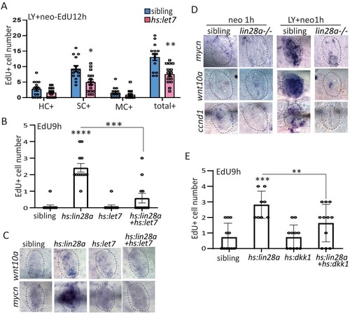
(A) We created hs:let7 transgenic line and found that it recapitulates the phenotype of lin28a-/- by decreasing proliferative SCs post LY+neo. (B) The induction of EdU+ proliferative cells was blocked by hs:let7, indicating that let7 microRNA acts downstream of lin28a to induce proliferation. (C) In situ hybridization results showed that expression of Wnt pathway genes, such as wnt10a and mycn, were increased in hs:lin28a. The activation of Wnt pathway genes in hs:lin28a were blocked when let7 was overexpressed, indicating that let7 acts downstream of lin28a to inhibit Wnt pathway. (D) Expressions of Wnt pathway genes, such as mycn, wnt10a and ccnd1 (cyclind1), were highly induced at 1 hr post LY+neo in sibling, but were not detected in lin28a-/-. (E) Inhibition of Wnt pathway with hs:dkk1 decreased lin28a-induced EdU+ proliferative cells, indicating that Wnt activation acts downstream of lin28a to induce regeneration.

Notch and Fgf pathways were not affected by <italic>lin28a</italic> overexpression.

In situ results show that expression of her4 and dkk2, Notch pathway genes, and fgf3, a Fgf pathway gene, were not affected in hs:lin28a post neo.

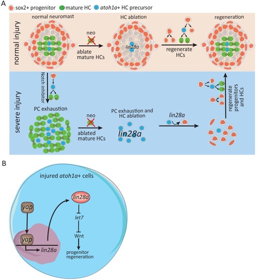
Schematic model illustrates how Yap-lin28a pathway regulates progenitor regeneration post severe injury.

(A) It’s known that sox2+ progenitors that are preserved post neo-induced HC injury divide and differentiate to regenerate HCs. To simulate the situation in mammalian inner ear where the progenitors are absent, we created a severe injury model by exhausting sox2+ progenitors with Notch inhibitor and ablating mature HCs with neomycin. We found that the exhausted sox2+ progenitors in severe injury have high potential to restore themselves within 48 hr, with HCs being regenerated afterwards. The atoh1a+ HC precursors, the main population that survived post severe injury, were converted into proliferative progenitors to initiate regeneration through yap-lin28a pathway. (B) Yap is activated in atoh1a+ HC precursors post severe injury and binds directly to lin28a promoter for initiating its transcription. Lin28a activates Wnt pathway through microRNA let7 to promote regenerative proliferation.

Acknowledgments
This image is the copyrighted work of the attributed author or publisher, and ZFIN has permission only to display this image to its users. Additional permissions should be obtained from the applicable author or publisher of the image. Full text @ Elife