- Title
-
Hedgehog signaling is required at multiple stages of zebrafish tooth development
- Authors
- Jackman, W.R., Yoo, J.J., and Stock, D.W.
- Source
- Full text @ BMC Dev. Biol.
|
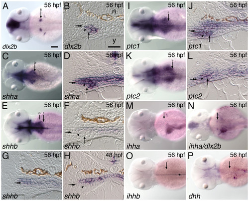
Hedgehog pathway expression during zebrafish tooth development. The hedgehog ligand shha and receptors ptc1 and ptc2 are expressed in the developing zebrafish pharyngeal dentition but we do not detect tooth-related expression of the ligands shhb, dhh, ihha, or ihhb. (A, C, E, I, K, M-P) Dorsal views of 56 hpf embryos, anterior to the left, with the location of the right-side pharyngeal tooth germ and anterior/posterior position of adjacent section indicated by an arrow. (B, D, F, H, J, L) Transverse sections of 56 hpf embryos at the level of the developing pharyngeal teeth. The lumen of the pharynx is collapsed in these histological preparations, leaving no space between the upper and lower epithelial layers. The lower pharyngeal epithelium (double arrow), dental epithelium (arrowhead), dental mesenchyme (arrow), notochord (n), and yolk (y), are indicated. (A, B) dlx2b, a discrete marker of zebrafish pharyngeal tooth germs, is expressed in both dental epithelium and mesenchyme [42]. Only epithelial expression is visible in B, however. (C, D) The hedgehog ligand shha is expressed widely in the pharyngeal epithelium including the dental epithelium, but not in the dental mesenchyme. (E, F) In contrast, at the anterior-posterior level of the developing teeth, shhb expression appears restricted to the upper pharyngeal epithelium and is absent from the lower epithelium and any part of the tooth germs. (G) A more rostral section from the same specimen as (F) exhibits expression in both pharyngeal epithelial layers. (H) Expression of shhb is also not seen in tooth germs at other stages of development such as 48 hpf when dental epithelial thickening is observed. The hedgehog receptors ptc1 (I, J) and ptc2 (K, L) are expressed widely in both the pharyngeal epithelium and in adjacent mesenchyme, encompassing both tissue layers in the tooth germs. We searched for, but were unable to detect, dental expression of the hedgehog ligands ihha (M; N, double label with dlx2b), ihhb (O; asterisk: notochord expression), and dhh (P). Scale bars: (A) 100 μm, (B) 25 μm. EXPRESSION / LABELING:
|
|
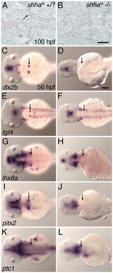
Zebrafish mutant for shha do not exhibit tooth-related developmental gene expression and do not form mature teeth. (A, B) Ventral views of the pharyngeal region at 100 hpf stained with alizarin red to highlight calcified structures. Teeth appeared normal in wild-type and shhat4 heterozygous individuals (A), but were not detected in shhat4 homozygous mutant siblings (B). mRNA in situ hybridization analysis at 56 hpf demonstrated that pharyngeal tooth-related expression of dlx2b (C, D), fgf4 (E, F), lhx8a (G, H), and pitx2 (I, J) was completely absent from homozygous mutant embryos but present in sibling controls possessing at least one wild type shha allele. Expression of ptc1 in the tooth-forming region appeared to be maintained in all genotypes (K, L). (C-L) Dorsal views, anterior to the left, position of right side tooth germs indicated (arrows). Scale bars: (B) 50 μm, (D) 100 μm. |
|
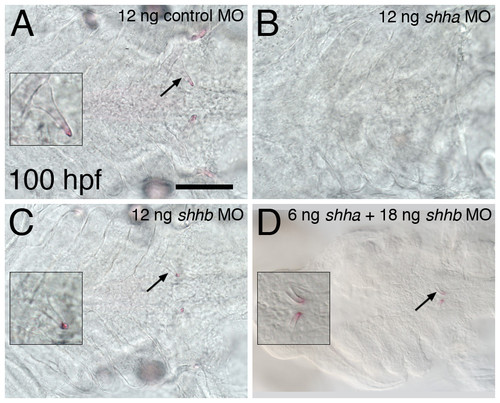
Antisense RNA inhibition of shha, but not shhb, prevents the formation of mature teeth. Ventral views of the tooth-forming region of alizarin red stained 100 hpf embryos. Teeth develop normally in control morpholino injected embryos (A, left-side tooth shown in inset). Teeth are eliminated from shha MO injected embryos (B), but retained in embryos injected with an identical amount (12 ng) of the shhb morpholino (C). The combination of 6 ng shha MO with 18 ng shhb MO results in teeth forming closer to the midline and appearing slightly developmentally delayed but otherwise with normal morphology (D). Scale bar 100 μm. PHENOTYPE:
|
|
Hedgehog requirements throughout tooth development. Inhibition of hedgehog signaling with cyclopamine (CyA) starting at 30 hpf prevents tooth morphogenesis, and treatments at subsequent stages reveal later hedgehog signaling requirements during tooth formation. (A-E) mRNA in situ hybridizations of pharyngeal tooth gene expression at 56 hpf for ptc1 (A), dlx2b (B), fgf4 (C), lhx8a (D), and pitx2 (E) in control embryos exposed to 0.5% EtOH from 30-56 hpf (dorsal views, anterior to the left, right side tooth germs indicated with an arrow, asterisks designate pectoral fin or girdle expression). (G-K) Embryos exposed to 50 μM CyA and 0.5% EtOH from 30-56 hpf show severely reduced or absent pharyngeal tooth expression of ptc1 (G), dlx2b (H), fgf4 (I), and lhx8a (L); but pitx2 (K) expression is maintained. (F, L) Transverse sections through the pharyngeal tooth forming region of 56 hpf embryos treated from 30 to 56 hpf in 0.5% EtOH with and without 50 μM CyA. Dental epithelial morphogenesis is highlighted by pitx2 mRNA expression in control embryos (F, arrow), but after CyA exposure the lower margin of the pharyngeal epithelium lacks the thickening or curved appearance characteristic of early tooth morphogenesis (L, arrow). (M-R) Right-side first pharyngeal tooth (arrows) in CyA or control treated, alcian blue stained and cleared 100 hpf larvae. No mature tooth formation was visible when CyA treatment was begun by 36 hpf (M). CyA exposure from 47 hpf allowed some limited mineralized morphogenesis, but it severely disrupted the shape of the entire tooth, causing it to appear unorganized (N). Treatment from 52 hpf resulted in teeth with a more regular appearance, but somewhat small and rounded (O). 60 hpf (P), and 72 hpf (Q), treatment resulted in teeth with only the later-developing shaft and base of the tooth having an abnormal rounded morphology (arrowhead) relative to controls (R). Scale bars: (A) 100 μm, (F) and (R) 25 μm. EXPRESSION / LABELING:
|
|
Overexpression of shha does not alter tooth formation or morphology. (A-F) Control shha overexpression experiments. After heat-shock at 12 hpf, 65 hpf control embryos display normally-developing retinas (A, arrow) whereas the ventral retinas of shha:GFP injected siblings is severely reduced (B, arrow). While absent from controls, GFP expression is visible in shha:GFP injected embryos, including in the region where teeth are forming (B, arrowhead), and appears to be concentrated in extracellular spaces. (C) ptc1 mRNA expression in uninjected embryos at 18 hpf subsequent to a heat shock at 12 hpf. (D) Embryos injected with the shha:GFP construct undergoing the same heat shock treatment exhibit strong upregulation of ptc1. Similarly, relative to controls (E), shha:GFP injected embryos showed increased ptc1 expression at 42 hpf after heat shock at 36 hpf (F). However, shha overexpression at neither stage altered mineralized tooth shape (arrow) or timing of tooth development as visualized at 100 hpf (G, H). Scale bar 25 μm. |


