
Fig. 6
- ID
- ZDB-IMAGE-241119-26
- Genes
- Publication
- England et al., 2024 - Transcriptional regulators with broad expression in the zebrafish spinal cord
- All Figures
- Figures for England et al., 2024
|
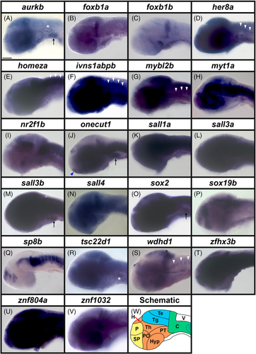
Fig. 6 Transcription factor and transcriptional regulator gene expression in zebrafish brain at 36 h. (A–V) Lateral views of heads in WT zebrafish embryos at 36 h. A minimum of five embryos were analyzed per gene (see Experimental Procedures for further details). (W) Schematic of a lateral view of the head of a 36 h zebrafish embryo. The telencephalon (dorsal forebrain, yellow) consists dorsally and ventrally of the pallium (P) and subpallium (SP) respectively. The diencephalon (ventral forebrain, orange) consists of the habenula (H), the hypothalamus (Hyp), the thalamic region (Th), the posterior tuberculum (PT) and the preoptic region (PO). The midbrain (blue) consists dorsally and ventrally of the tectum (Te) and the tegmentum (Tg), respectively. The hindbrain (green) consists of the cerebellum (C). V, ventricle in the hindbrain. Black dotted line = midbrain-hindbrain boundary. Blue dotted lines = otic vesicle. Transcriptional regulator gene (A) aurkb, and transcription factor genes (B) foxb1a, (C) foxb1b, (D) her8a, (E) homeza, (F) ivns1abpb, (G) mybl2b, (H) myt1a, (I) nr2f1b, (J) onecut1, (K) sall1a, (L) sall3a, (M) sall3b, (N) sall4, (O) sox2 (P), sox19b, (Q) sp8b, (R) tsc22d1, (S) wdhd1, (T) zfhx3b, (U) znf804a, and (V) znf1032 are all variably expressed in the brain. (D, E, F, G, S) White arrowheads depict weak expression in the rhomobomeres of the hindbrain. (A, R) aurkb and tsc22d1 are expressed in the otic vesicles (white asterisks). (A, J, M, O) aurkb, onecut1, sall3b and sox2 are expressed in the branchial (gill) mesenchyme (black arrow). (J and data not shown) onecut1 and sp8b are expressed in the olfactory bulbs (blue arrowhead). (N) sall4 in situ hybridization experiments were performed with the molecular crowding reagent Dextran Sulfate (see Experimental Procedures for rationale). All other in situ hybridization experiments in this figure were performed without this reagent. Scale bar: 100 μm.