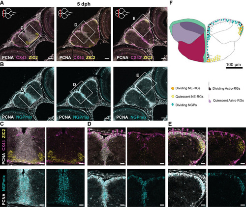
Progenitor heterogeneity in the developing telencephalon. (A,B) Adjacent coronal sections of the telencephalon at 5 dph along the anterior-posterior axis. The level of each section is indicated with a red line on the top-view brain illustration in the upper-left corner of each panel in A. HCR-FISH targeting CX43 (pan Astro-RG, magenta) and ZIC2 (NE-RG, yellow) (A) or NGPmix (turquoise) (B), is combined with immunohistochemical staining for the proliferation marker PCNA (white). NGPs account for the bulk of proliferation; Astro-RGs and NE-RGs appear both dividing and non-dividing depending on the location in the telencephalon. (C-E) Magnifications of the regions in the boxes in A and B. Non-dividing Astro-RGs (CX43+, PCNA−) are indicated with a magenta arrowhead and appear isolated in between stretches of dividing NGPs. (F) Illustration of a 5 dph coronal telencephalic section at mid-anterior-posterior level. The distribution of dividing and non-dividing Astro-RGs, NE-RGs and NGPs is displayed in the ventricular zone. Scale bars: 50 µm (A,B); 20 µm (C-E).
|
