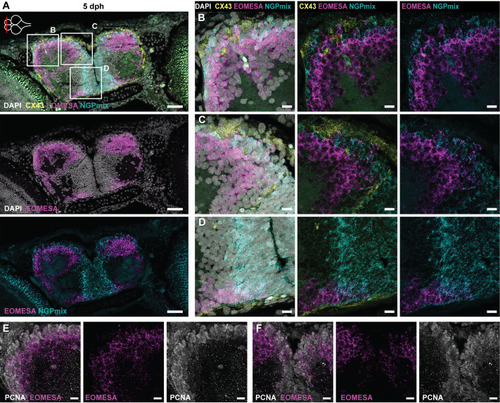
Intermediate progenitors in the killifish pallium. (A) HCR-FISH targeting CX43 (Astro-RGs, yellow), NGPmix (turquoise) and the immature excitatory neuron marker EOMESA (magenta) in combination with the nuclear stain DAPI (white) on a coronal section of the telencephalon at 5 dph. The anterior-posterior level is depicted with a red line on the top-view brain illustration in the upper-left corner. (B-D) Magnifications of the regions in the boxes in A. Intermediate progenitors (EOMESA+/NGPmix+) are found at the pallial neurogenic region and ventral subpallium, dispersed in between Astro-RGs (only in pallium) and NGPs. (E,F) HCR-FISH targeting EOMESA (magenta) in combination with immunohistochemical staining for the proliferation marker PCNA (white). The panels are magnifications of zones on a section of a comparable anterior-posterior level as in A. E and F are magnifications of the lateral pallium and dorsal midline, respectively. Proliferating PCNA+/EOMESA+ progenitors are present at the midline and pallial surface. Scale bars: 50 µm (A); 10 µm (B-F).
|
