|
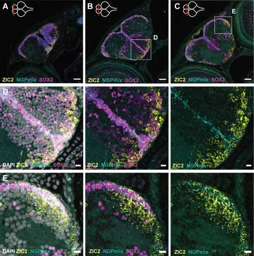
Distribution of the non-glial and neuroepithelial-like progenitors. (A-C) HCR-FISH targeting NGPmix (STMN1A+HMGB2A, NGPs, turquoise) and ZIC2 (NE-RGs, yellow) in combination with immunohistochemical staining for the progenitor marker SOX2 (magenta) on coronal sections along the anterior-posterior axis in the 5 dph telencephalon. (D,E) Magnifications of the regions in the boxes in B and C. The HCR-FISH targeting ZIC2 (yellow) and NGPmix (turquoise) and SOX2 staining (magenta) are combined with the nuclear stain DAPI (white). NGPs and NE-RGs are present in the subpallial midline and posterior pallial neurogenic regions. In the subpallium, both cell types retain their progenitor profile (SOX2+), while in the pallium, NE-RG3 loses its progenitor profile (SOX2−) in the parenchyme. Scale bars: 50 µm (A-C); 10 µm (D,E).
|