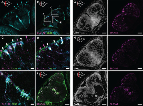
Radial glia patterns in the developing telencephalon. (A) Immunohistochemical staining of GS (turquoise) on a 5 dph telencephalon section at a posterior level. Isolated GS+ RGs are positioned one by one at the pallial surface (turquoise arrowheads) and in a dense cluster at the subpallial midline. (B) HCR-FISH targeting SLC1A2 (magenta) and CX43 (green) in combination with the immunohistochemical GS (turquoise) staining on a coronal section at the mid-anterior-posterior level. (C-E) Magnification of the squares in B. GS+/SLC1A2+/CX43+ RGs (white arrowheads) are interspersed with SLC1A2+-only RGs (magenta arrowheads), which are most probably not fully mature yet, at the pallial surface. (F) HCR-FISH targeting SLC1A2 (magenta) and CX43 (green) on a coronal section of the posterior telencephalon at 5 dph. At the midline, a dense cluster of CX43+SLC1A2+ RGs could be identified as the astroglia subcluster Astro-RG2. (G-I) Coronal sections of the telencephalon at 5 dph along the anterior-posterior axis. Left panels show the nuclear stain DAPI (white) to delineate the nuclear composition and density of the telencephalic domains. Right panels display HCR-FISH targeting SLC1A2 (magenta). Cell bodies, at the ventricular surface, and fiber structures, running towards the pia, are visible on all levels. The anterior-posterior position of the sections is indicated with a red line on a top-view illustration of the brain in the upper-left corner of the panels. Scale bars: 50 µm (A,B,F-I); 10 µm (C-E). AC, anterior commissure; HCR-FISH, hybridization chain reaction–fluorescent in situ hybridization.
|
