IMAGE

Fig. 6.

ID
ZDB-IMAGE-250502-18
Source
Figures for Zandecki et al., 2025
Image
Figure Caption

Fig. 6.

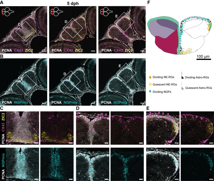
Progenitor heterogeneity in the developing telencephalon. (A,B) Adjacent coronal sections of the telencephalon at 5 dph along the anterior-posterior axis. The level of each section is indicated with a red line on the top-view brain illustration in the upper-left corner of each panel in A. HCR-FISH targeting CX43 (pan Astro-RG, magenta) and ZIC2 (NE-RG, yellow) (A) or NGPmix (turquoise) (B), is combined with immunohistochemical staining for the proliferation marker PCNA (white). NGPs account for the bulk of proliferation; Astro-RGs and NE-RGs appear both dividing and non-dividing depending on the location in the telencephalon. (C-E) Magnifications of the regions in the boxes in A and B. Non-dividing Astro-RGs (CX43+, PCNA) are indicated with a magenta arrowhead and appear isolated in between stretches of dividing NGPs. (F) Illustration of a 5 dph coronal telencephalic section at mid-anterior-posterior level. The distribution of dividing and non-dividing Astro-RGs, NE-RGs and NGPs is displayed in the ventricular zone. Scale bars: 50 µm (A,B); 20 µm (C-E).

Acknowledgments
This image is the copyrighted work of the attributed author or publisher, and ZFIN has permission only to display this image to its users. Additional permissions should be obtained from the applicable author or publisher of the image. Full text @ Biol. Open