|
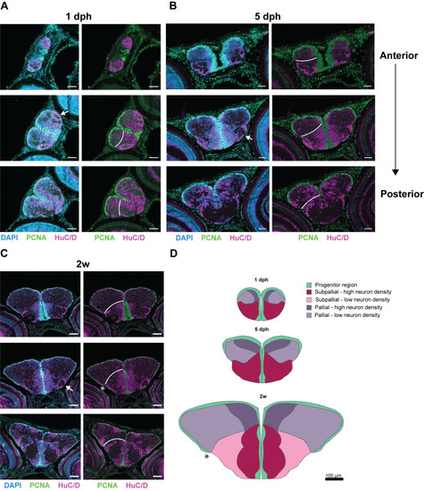
Explosive growth of dorsal and ventral telencephalic domains. (A-C) Immunohistochemical staining for PCNA (green) and HuC/D (magenta) in combination with a nuclear stain (DAPI, blue) in coronal brain sections. Three levels of the anterior-posterior axis – anterior-mid-posterior – are shown for killifish at 1 dph (A), 5 dph (B) and 2 weeks (C), from anterior to posterior. White lines represent the pallial-subpallial border. (D) On scale illustration summarizing the distribution of progenitor (green) and neuronal regions (shades of red/pink/purple) on coronal telencephalic sections of juveniles at 1 dph, 5 dph, and 2 weeks. Example sections of a comparable anterior-posterior level were selected based on the presence of a specific ventrolateral neuronal cluster (arrows, A-C) and the progenitor region extending from the dorsal to the ventral surface (dashed lines, A-C). (A-B) Scale bars: 50 µm (A,B); 100 µm (C,D). dph, days post-hatching; w, week.
|