|
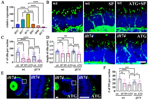
Maternal Ift74 contributes to the slow photoreceptor degeneration. (A) qRT-PCR results showing relative expression level of ift-b genes in one-cell stage embryos. The expression of the ift74 gene was set as 100%. (B) Confocal images showing photoreceptor connecting cilia in the retinae of 5 dpf wild-type, ift74 morphants, and mutant larvae. Cilia were stained with anti-acetylated alpha tubulin antibody (green). (C,D) Dot plots showing the number and length of connecting cilia in different groups as indicated. (E) Confocal images showing rod outer segments visualized with zpr-3 antibody in 5 dpf ift74 mutants injected with control or ATG morpholino as indicated. Enlarged views of the boxed area are shown on the right. (F) Statistical results showing the number of outer segments per retina section in different groups as indicated. Scale bars: B, 5 µm; E, 30 µm. * p < 0.05, ** p < 0.01, *** p < 0.001, **** p < 0.0001, ns, no significant.
|