
FIGURE
Figure 4
Figure 4
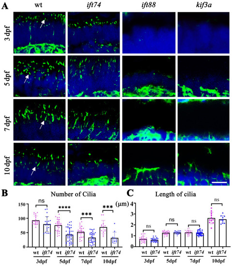
Photoreceptor connecting cilia in ift74, ift88, and kfi3a mutants. (A) Confocal images showing photoreceptor connecting cilia in the retinae of wild-type and mutant larvae at different stages as indicated. Cilia were stained with anti-acetylated alpha tubulin antibody (green). Arrows indicate the connecting cilia. (B,C) Dot plots showing the number and length of connecting cilia in different groups as indicated. Scale bar: 10 µm. *** p < 0.001, **** p < 0.0001, ns, no significant. |
Expression Data
| Antibody: | |
|---|---|
| Fish: | |
| Anatomical Term: | |
| Stage Range: | Protruding-mouth to Days 7-13 |
Expression Detail
Antibody Labeling
Phenotype Data
| Fish: | |
|---|---|
| Observed In: | |
| Stage Range: | Protruding-mouth to Days 7-13 |
Phenotype Detail
Acknowledgments
This image is the copyrighted work of the attributed author or publisher, and
ZFIN has permission only to display this image to its users.
Additional permissions should be obtained from the applicable author or publisher of the image.
Full text @ Int. J. Mol. Sci.