|
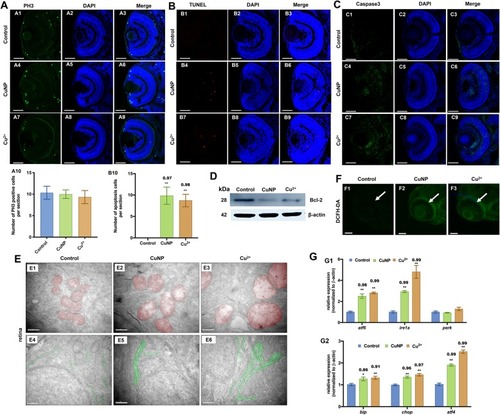
Cell proliferation, apoptosis, and oxidative & ER stresses in copper-stressed embryos. A Cell proliferation assay by PH3 staining (green dots). B Cell apoptosis assay by TUNEL (red dots) detection. (A, B) sections of embryonic eyes. A10, number of PH3 positive cells per section, B10, average number of apoptotic cells of retinal sections in each group (n > 3, 3–5 sections from each embryo were used for counting the red positive apoptotic cells). C Immunostaining of Caspase3 (green) in retina sections. D Western blot detection with antibody Bcl-2 in copper-stressed embryos. E TEM analysis of retinal cells in copper-stressed embryos at 96 hpf. E1-E6: sagittal slides of retina, red color indicating mitochondria and green color indicating ER. F DCHF-DA assay of embryonic retina. G qRT-PCR detection of ER-stressed genes in embryos at 96 hpf. Scale bars: A1-A9, B1-B9 and C1-C9, 50 μm; E1-E6, 0.5 μm; F1-F3, 100 μm; **, P < 0.01; *, P < 0.05
|