
Fig. 3
- ID
- ZDB-IMAGE-200406-150
- Genes
- Antibodies
- Publication
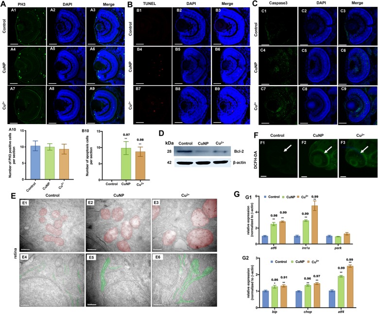
- Zhao et al., 2020 - Copper induce zebrafish retinal developmental defects via triggering stresses and apoptosis
- All Figures
- Figures for Zhao et al., 2020
|
Fig. 3
Cell proliferation, apoptosis, and oxidative & ER stresses in copper-stressed embryos.