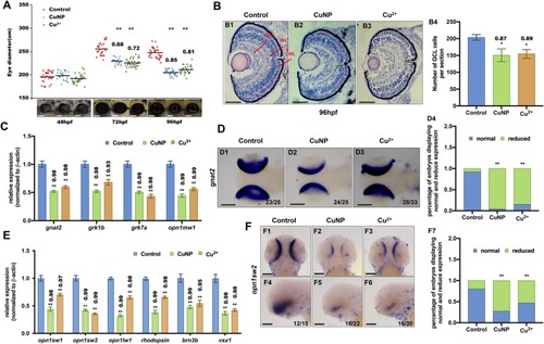
Retinal developmental defects in copper stressed embryos. A Measurement of eye diameter of the embryos from control, Cu2+- and CuNPs- stressed groups at 48 hpf, 72 hpf, and 96 hpf, respectively. B H&E staining analysis of retina of embryos from control (B1), Cu2+-stressed (B2) and CuNPs-stressed groups (B3) at 96 hpf. Control embryos at 96 hpf developed normal retina with a differentiated GCL (ganglion cell layer), an INL (inner nuclear layer), and an ONL (outer nuclear layer) (indicated by the red arrows). (B4) Average number of GCL cells per section/per embryo in each group (n > 3, 3–5 sections from each embryo were used for counting the GCL cells). C Expression of retinal marker genes gnat2, grk1b, grk7a, and opnlmw1 in embryos from control, Cu2+-stressed, and CuNPs-stressed groups. D WISH data of gnat2 in embryos from control, Cu2+-stressed, and CuNPs-stressed groups, respectively (D1-D3), and the percentage of embryos exhibiting reduced expression in different groups (D4). E Expression of retinal marker genes opn1sw1, opn1sw2, opn1lw1, rhodopsin, brn3b, and vsx1 in embryos from control, Cu2+ − stressed, and CuNPs-stressed groups. F WISH data of opn1sw2 in embryos from control, Cu2+-stressed, and CuNPs-stressed groups, respectively (F1-F6), and the percentage of embryos exhibiting reduced expression in different groups (F7). B1-B3, sagittal slides in eyes domain; D1-D3, dorsal view, anterior to the left; F1-F3, dorsal view, anterior to the up; F4-F6 lateral view, anterior to the left. Scale bar: A, B1-B3, and F1-F6, 100 μm; D1-D3, 200 μm **, P < 0.01; *, P < 0.05
|

